Download
1 / 22
220 likes | 557 Views
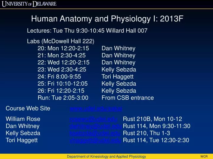
Human Anatomy and Physiology I: 2013F Lectures: Tue Thu 9:30-10:45 Willard Hall 007 Labs (McDowell Hall 222) 20: Mon 12:20-2:15 Dan Whitney 21: Mon 2:30-4:25 Dan Whitney 22: Wed 12:20-2:15 Dan Whitney 23: Wed 2:30-4:25 Kelly Sebzda 24: Fri 8:00-9:55 Tori Haggett

E N D
Human Anatomy and Physiology I: 2013F • Lectures: Tue Thu 9:30-10:45 Willard Hall 007 • Labs (McDowell Hall 222) • 20: Mon 12:20-2:15 Dan Whitney • 21: Mon 2:30-4:25 Dan Whitney • 22: Wed 12:20-2:15 Dan Whitney • 23: Wed 2:30-4:25 Kelly Sebzda • 24: Fri 8:00-9:55 ToriHaggett • 25: Fri 10:10-12:05 Kelly Sebzda • 26: Fri 12:20-2:15 Kelly Sebzda • Run: Tue 2:05-3:00 From CSB entrance • Course Web Site www.udel.edu/sakai • William Rose rosewc@udel.edu Rust 210B, Mon 10-12 • Dan Whitney dwhitney@udel.edu Rust 114, Mon 9:30-11:30 • Kelly Sebzdaksebzda@udel.edu Rust 210, Thu 1-3 • Tori Haggettvhaggett@udel.edu Rust 114, Tue 12:30-2:30 Department of Kinesiology and Applied Physiology
A. & P. I: 2013F Instructors Dan Whitney Labs 20, 21, 22 Physiology of bone and adipose tissue Exercise Science, Univ. of New Hampshire Kelly Sebzda Labs 23, 25, 26 Exercise to prevent chronic illness (cancer, heart disease) Kinesiology (Exercise & Sports Science), Temple Univ. Tori Haggett Lab 24 Rehabilitation biomecahnics and gait analysis Exercise Science, Furman University William Rose Lectures Cardiovascular physiology, biomechanics, computer simulations in physiology Department of Kinesiology and Applied Physiology
Pre-clinical Anatomy and Physiology I KAAP309-13F Grading – see syllabus. 70% Classroom 65%: Eleven tests (worst is dropped) 5%: Clicker 30% Laboratory 18% Group: Three simulation labs, two group projects, peer grade 12% Individual: Four lab practical exams Department of Kinesiology and Applied Physiology
Pre-clinical Anatomy and Physiology I KAAP309-13F UD Capture: Recording of what is projected on screen and classroom audio. http://udcapture.udel.edu/2013f/kaap309-010/ Clickers: Register your clicker on Sakai. Clicker questions: 1 point for answering, 1 more point if correct. Clicker grade: Full credit if you get 75% or more of the points available. Reduced proportionally if not. No adjustments for forgotten or broken clickers, low batteries, etc. If a student is observed using more than one clicker, both clicker numbers will be noted and grades reduced for both students. Department of Kinesiology and Applied Physiology
“The moment one gives close attention to anything, even a blade of grass, it becomes a mysterious, awesome, indescribably magnificent world in itself.” — Henry Miller
A single word embodies the entire foundation of Western medicine. Its three letters set the tone for a distinctive world-view of healing and for the science upon which it is based... That word is see. Since Western medicine’s origins in ancient Greece some twenty-five hundred years ago, the perspective of its researchers and practitioners has been that the processes of both normal and diseased physiology must be visualizable in order to be understood in any realistic way. It is necessary, in other words, to foster a system of comprehension in which at least the mind’s eye but preferably the literal eye faithfully sees the body’s components as they are actually functioning… The Western doctor of today should be able to draw a picture of his patient’s organs, tissues, and even cells, depicting the events that are happening within them... The aim of medicine is to describe and document every step of the process by which health becomes sickness and is then restored. The history of Western medicine has been the history of its disciples at first gradually and most recently at dizzying speed uncovering in ever more minute detail the steps within those steps. The Mysteries Within. Sherwin Nuland, 2000. See …\reserve\nuland_on_seeing.doc for longer excerpt.
A&P in the News • N.Y. Times: • “Weight index doesn’t tell the whole truth” • How measure thinness/fatness? • (1 , 2) • http://www.nytimes.com/2010/08/31/health/31brod.html?ref=health • What is Mr Olympia’s BMI? • <18.5 (underweight) • 18.5-24.9 (normal) • 25-29.9 (overweight) • 30-40 (obese) • >40 (morbidly obese) Department of Kinesiology and Applied Physiology
Planes of section, directional terms, and body regions will be discussed in the lab. Department of Kinesiology and Applied Physiology
Figure 1.9 2 Frontal plane Sagittal plane Transverse plane The major sectional planes
Figure 1.9 1 Superior Left Right Cranial Proximal Posterior or dorsal Anterior or ventral Lateral Medial Caudal Proximal Distal The principal directional terms Distal Inferior
Figure 1.8 1 Nasus or nose (nasal) Frons or forehead (frontal) Oculus or eye (orbital or ocular) Cranium or skull (cranial) Cephalon or head (cephalic) Auris or ear (otic) Cephalon or head (cephalic) Facies or face (facial) Bucca or cheek (buccal) Cervicis or neck (cervical) Acromion (acromial) Cervicis or neck (cervical) Dorsum or back (dorsal) Oris or mouth (oral) Mentum or chin (mental) Thoracis or thorax, chest (thoracic) Axilla or armpit (axillary) Mamma or breast (mammary) Brachium or arm (brachial) Olecranon or back of elbow (olecranal) Trunk Upper limb Abdomen (abdominal) Antecubitis or front of elbow (antecubital) Umbilicus or navel (umbilical) Lumbus or loin (lumbar) Antebrachium or forearm (antebrachial) Pelvis (pelvic) Carpus or wrist (carpal) Palma or palm (palmar) Palma or palm (palmar) Manus or hand (manual) Gluteus or buttock (gluteal) Pollex or thumb Lower limb Digits or phalanges or fingers (digital or phalangeal) Inguen or groin (inguinal) Popliteus or back of knee (popliteal) Pubis (pubic) Patella or kneecap (patellar) Femur or thigh (femoral) Crus or leg (crural) Sura or calf (sural) Tarsus or ankle (tarsal) Calcaneus or heel of foot (calcaneal) Digits or phalanges or toes (digital or phalangeal) Pes or foot (pedal) Planta or sole of foot (plantar) Hallux or great toe The anatomical position in anterior view The anatomical position in posterior view
Figure 1.10 2 – 4 THORACIC CAVITY Each lung is enclosed within a pleural cavity, lined by a shiny, slippery serous membrane called the pleura (PLOO-ra). Note the orientation of the section. Unless otherwise noted, all cross sections are shown as if the viewer were standing at the feet of a supine person and looking toward the head. Heart in pericardial cavity Right lung in right pleural cavity Left lung in left pleural cavity The body cavities: the thoracic cavity and the abdominopelvic cavity A horizontal section through the thoracic cavity shows the relationship between the subdivisions of the ventral body cavity in this region. The pericardial cavity is embedded within the mediastinum, a mass of connective tissue that separates the two pleural cavities and stabilizes the positions of embedded organs and blood vessels. BODY CAVITIES ABDOMINOPELVIC CAVITY During development, the portion of the original ventral body cavity extending into the abdominopelvic cavity remains intact as the peritoneal (per-i-tō-NĒ-al) cavity, a chamber lined by a serous membrane known as the peritoneum (per-i-tō-NĒ-um). A few organs, such as the kidneys and pancreas, lie between the peritoneal lining and the muscular wall of the abdominal cavity. Those organs are said to be retroperitoneal (re-trō- per-i-tō-NĒ-al; retro, behind). Diaphragm Peritoneum (red) showing the boundaries of the peritoneal cavity The abdominal cavity contains many digestive glands and organs THORACIC CAVITY Retroperitoneal area The pelvic cavity contains the urinary bladder, reproductive organs, and the last portion of the digestive tract; many of these structures lie posterior to, or inferior to, the peritoneal cavity. The diaphragm, a muscular sheet, separates the thoracic cavity from the abdominopelvic cavity. ABDOMINOPELVIC CAVITY
Serous membranes (serosa): line body cavities. Parietal & visceral. Department of Health, Nutrition, and Exercise Sciences