Download

1 / 59
820 likes | 2.07k Views
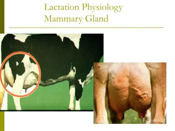
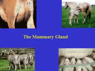
Lactation Physiology Mammary Gland. Prevention. Healthy cows with good immune systems will be able to fight off mastitis infections. Many mastitis pathogens are eliminated by the cow's own defense system. Assure that the diets are balanced for Vitamin E and Selenium.

E N D
Prevention • Healthy cows with good immune systems will be able to fight off mastitis infections. Many mastitis pathogens are eliminated by the cow's own defense system. • Assure that the diets are balanced for Vitamin E and Selenium. • Immunization with J5 vaccine will not prevent infection but will decrease the severity of clinical signs.
The mammary gland nourishes the neonate • Exocrine gland; common to all mammals • Function: nourish the neonate • Food source: fat, protein, sugar (CHO), vitamins, minerals, water • Protection: immunoglobulins (first Ab protection; absorbed via intestinal tract)
The mammary gland is part the reproductive system • The mammary gland is loosely considered part of the reproductive system: • Serves a “reproductive function”; nourishment of the neonate = survival of species. • Relies on same endocrine (hormonal) support for development and function. Example: gonadal steroids, prolactin, etc.
Udder consists of four separate glands A teat hangs from each quarter Bottom of teat closed by sphincter muscle known as streak canal Can have extra nonfunctional teats Called supernumerary teats Removed when calf is young Mammary Gland Structure
Mammary Gland Structure • Conformation of teats • Vary in shape from cylindrical to conical • Rear teats are usually shorter • Each teat has one streak canal • Teats should be moderately sized and located centrally on each quarter • Sphincter in each teat should be tight enough to prevent leakage • Teats are hairless
Mammary Gland Structure • Support system = Stroma (connective tissue) • Glandular; secreting tissue = Parenchyma • Alveoli- secreting epithelial cells • Duct system- lined by epithelial cells • Lobules & lobes- clusters of alveolar tissue supported by connective tissue
Separate Mammary Glands-Quarters 40% 60% Front quarter is smaller
Mammary Gland Structure/Suspension • Intermammary groove separates left and right halves of the udder • Udder can weigh anywhere from 7 to 165 pounds • May support up to 80 pounds of milk • Rear quarters secrete 60% of the milk • Udder continues to grow in size until cow is 6 years of age • Well attached udder fits snugly against the abdominal wall in front and on the sides • Extends high between thighs in rear • 3 major supporting structures • Skin • Median suspensory ligament • Lateral suspensory ligament
Mammary Gland Suspension • Skin • Minor role in support • Median suspensory ligament • Separates right and left halves of udder • Connects udder to abdominal wall • Lamellae • Elastic tissue which responds to weight of milk in udder • Lateral suspensory ligament • Inflexible • Surround the outer wall of udder • Attached to prepubic and subpubic tendons • Intermammary groove formed where lateral suspensory ligament and median suspensory ligament meets
Fig 29-3. An illustrated view of the ligaments that permit udder suspension (Courtesy of Iowa State University)
Mammary Gland Support Medial suspensory ligament
Internal Anatomy • Streak canal • Functions to keep milk in udder and bacteria out of udder • Teat cistern • Duct in teat with capacity of 30-45 milliliters • Separated from streak canal by folds of tissue called Furstenberg’s rosettes • Gland cistern • Separated from teat cistern by the cricoid fold • Holds up to 400 milliliters of milk • Collecting area for the mammary ducts • From this branches the mammary ducts
Fig 29-4. A dissected mammary gland showing the gland cistern, teat cistern and streak canal (Courtesy of Mark Kirkpatrick)
Alveoli and Duct System • Alveoli is the basic milk producing unit • Small bulb-shaped structure with hollow center • Lined with epithelial cells that secrete milk • Each cubic inch of udder tissue contains 1 million alveoli • Each alveoli surrounded by network of capillaries and myoepithelial cell • Contraction of myoepithelial cell stimulates milk ejection • Groups of alveoli empty into a duct forming a unit called a lobule • Several lobules create a lobe • Ducts of lobe empty into a galatophore, which empties into the gland cistern • Ducts provide storage area for milk and a means for transporting it outside • Lined by two layers of epithelium
Alveolar Products • Alveolus: • basic secretory unit; lined by epithelial cells which synthesize and/or secrete: • lipid - triglycerides & free fatty acids (FFA) • protein - caseins • lactose - disaccharide; osmoreactive molecule (draws water) • minerals & vitamins - Ca, P, K; Vits. A, B, C, D • water
Circulation • One gallon of milk requires 400 gallons of blood being passed through udder • Ratio may increase in low producing cows • Blood enters the udder through external pudic arteries • Blood exiting udder from veins at the base of udder blood can travel through two routes • Via external pudic veins • Via subcutaneous abdominal veins
Fig 29-6. Blood flow to and from the mammary gland determines milk producing capability of the cow (Courtesy of Iowa State University)
Mammary Venous Circle Cranial Mammary Vein
Mammary gland defense system • Smooth muscle sphincter surrounding the teat canal inhibits bacterial closure. Because the teat canal lumen remains dilated for up to 2 hours after milking, feed cows after milking to keep them on their feet. • Keratin, a waxy substance derived from the teat canal lining partially occludes the lumen of the teat canal and inhibits bacterial penetration. Only infuse the tip of intramammary infusion cannulas into the teat canal. • Somatic Cells are the most important natural defense mechanism to infection. Leukocytes (mostly PMN, polymorphonuclear neutrophils) function by phagocytosing and killing bacteria. They may reach in the millions. • Antibodies and other soluble factors in milk. They coat bacteria and enhance PMN engulfment. They also interfere with bacterial adhesion to tissues, reducing multiplication and neutralizing toxins.
Lymphatic System • Helps regulate proper fluid balance within udder and combat infection • Fluid drained from tissue only travels away from udder • Blood capillary pressure • Contraction of muscles surrounding the lymph vessels • Valves that prevent backflow of lymph • Mechanical action of breathing • Lymph travels from udder to the thoracic duct and empties into blood system • Flow rates of lymph depend on physiological status of the cow
Lymphatic System • Fluid enters the lymph system through open-ended vessels called lacteals
Lymphatic System- Edema • Edema: • low pressure, passive system fed by a high pressure vascular system! • this situation results in pooling of interstitial fluid if evacuation of lymph is impaired Example: tissue trauma; increased mammary blood flow at parturition
Alleviating Mammary Edema • Preparturient milking may be helpful • store colostrum from healthy cows to feed calves • Frequent milkout to reduce mammary pressure • Diuretics, corticoids to reduce swelling • Mammary massage, icing • work fluid towards supramammary lymph nodes • Reduce salt intake • Don’t feed too much, too early before calving
Foul smell and necrotic odor • Arcanobacterium pyogenes • (another)anaerobe • Watery milk, swollen udder • Coliforms • Watery and red, sick cow • Staph aureus: poor prognosis • Subclinical mastitis: contagious organisms:Staphylococcus aureus, • Streptococcus agalactia, Mycoplasma bovis • Clinical mastitis: • Environmental organisms: Strep. nonag. Group:50% • Coliform organisms (Eschericia coli, Klebsiella, Enterobacter sp., etc.) • 90% of the time
Follicles and New Corpus Luteum Mature Corpus Luteum Reproductive Tract Cow’s Reproductive Cycle
Reproductive hormones Estrogen: positive feedback Progesterone:: negative feedback
Day 0 is considered to be estrus. • Days 1-5 are metestrus. • Days 6-17 are diestrus. • Days 18-20 are proestrus.
1. Transmitter with pressure sensitive button on top 3. The receiver accepts the mounting activity signal from the transmitter Receiver ~1/2 mile range 2. Mounted onto cow’s rump using a patch and adhesive ))))))))))))))))))))))))))))))))) 4. Information is transferred to computer for processing by HeatWatch software. Heat Watch
Ovsynch • Ovsynch uses two hormones • PGF2α and GnRH • Ovsynch occurs in three stages • Day 0 - Stage 1: GnRH injection to create a new follicle • Day 7 - Stage 2: PGF2α injection to end the currnet estrus cycle and regress the corpus luteum • Day 9 – Stage 3: Second GnRH injection to cause the new follicle to ovulate and release the egg • All cows will ovulate 24-32 hours after the second GnRH injection • Day 10 – Insemination
CIDR - Source: ansci.wisc.edu • CIDRs (Controlled Internal Drug Release) are an intravaginal progesterone insert used in the beef cattle, dairy cattle, goat and sheep industries. • The progesterone is released at a controlled rate into the bloodstream after insertion. • In all species, CIDRs are used for the synchronization of estrus. • This can be highly beneficial in large herds because with the synchronization of estrus, groups of cows and heifers can be bred at the same time in a narrow window.