Download
1 / 30
300 likes | 374 Views
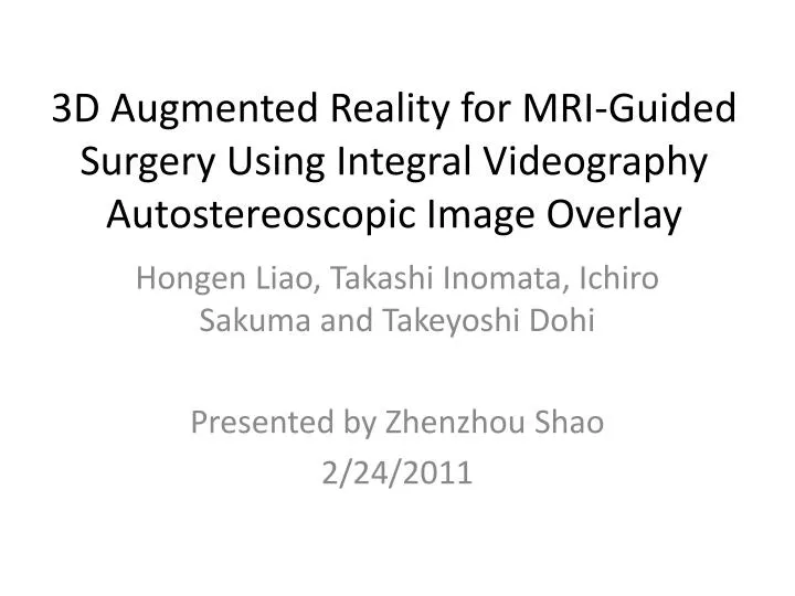
3D Augmented Reality for MRI-Guided Surgery Using Integral Videography Autostereoscopic Image Overlay. Hongen Liao, Takashi Inomata, Ichiro Sakuma and Takeyoshi Dohi Presented by Zhenzhou Shao 2/24/2011. Outline. Introduction Material and Methods System Configuration

E N D
3D Augmented Reality for MRI-Guided Surgery Using Integral VideographyAutostereoscopic Image Overlay Hongen Liao, Takashi Inomata, Ichiro Sakuma and Takeyoshi Dohi Presented by Zhenzhou Shao 2/24/2011
Outline • Introduction • Material and Methods • System Configuration • IV Image Display and Overlay Device • Registration of Spatial 3D Image in Patient • Software Alignment • Surgical Procedure • Experiment and Results • Conclusion
Introduction • Magnetic resonance imaging (MRI) • A medical imaging technique • Provides detailed information about soft tissue.
Introduction • Potential efficacy using MRI-guided surgery • Advantages • Enhance the surgeon’s capability • Decrease the invasiveness of surgical procedure • Increase the accuracy and safety • Disadvantages • Display of a set of 2D sectional images • Hand–eye coordination problem
Introduction • Augmented Reality (AR) • Superimpose the virtual model into the real scene. • Video see-through AR • Head mounted display (HMD) • Limited field of view • A lag for motion parallax • Cannot provide a natural view for multiple observers
Introduction • Optical see-through AR • Using a semi-transparent mirror for merging virtual model with a direct view. • Surgeon can see through the body. • Enhance the surgeon’s ability to perform a complex procedure. • Depth information is required.
Introduction • Integral Videography(IV)
Surgical Procedure • Calibrate the position of reflected IV image; • Place sterile fiducial markers on the surface of the patient’s body and scan the target area; • Segment the target of interest and markers from the MRI data. Perform patient-to-image registration to find the ;
Surgical Procedure • Render the IV images and transfer them to the overlay device; • Perform the surgical treatment under the guidance of IV image overlay; • After finishing the treatment, translate the patient into the scanner again and confirm surgical result.
Experiment and Results • Accuracy measurement • Implemented by using markers in a phantom simulating the human head.
Accuracy measurement • Five markers for registration and two for error measurement. • Marker: 10 mm in external diameter and 3 mm in internal diameter. • The distance between the center of the actual marker and that of the spatial projected IV marker was measured as an overlay error. • The mean value of the error was 0.90 mm, and the standard deviation was 0.21 mm
Targeting Experiment • Compare the procedure time and success rate of targeting an object using 2-D image guidance and IV overlay system guidance. • Phantom consisted of a plastic cube container filled with an agar.
Targeting Experiment • Six MRI markers were attached. • Three sets of acrylic cylinders with diameters of 1.5, 2 and 3 mm were embedded within the phantom.
Results of guidance 2-D image guidance IV overlay system guidance
Feasibility Evaluation • Evaluate the feasibility by a volunteer test. • Scan brain using MRI. • Motion parallax could be generated due to the motion of an observer. • The motion parallax of IV autostereoscopic brain images combined with the volunteer’s head was taken from various directions.
In Vivo Animal Experiment • Target a pig’s gallbladder. • A set of markers was attached to the skin of the surgical area.
In Vivo Animal Experiment • Surgical planning to minimizing the surgical exposure. • Surgical instrument is tracked. • The targeting experiment was performed by a medical doctor.
Conclusion • An autostereoscopic image overlay system for MRI-guided surgery is developed. • IV is employed to provide accurate 3-D spatial images and reproduces motion. • A fast and accurate spatial image registration method was developed. • Safe, easy, and accurate surgical diagnosis and therapy.