Download
1 / 4
40 likes | 63 Views
Today, computer aided system is widely used in various fields. Among them, the brain tumor detection is an important task in medical image processing. Early diagnosis of brain tumors plays an important role in improving treatment possibilities and increases the survival rate of the patients. Manual segmentation of brain tumors for cancer diagnosis, from large amount of Magnetic Resonance Imaging MRI images generated in clinical routine, is a difficult and time consuming task or even generates errors. So, the automatic brain tumor segmentation is needed to segment tumor. The purpose of the thesis is to detect the brain tumor quickly and accurately from the MRI brain image. In the system, the average filter is used to remove noise and make smooth an input MRI image and threshold segmentation is applied to segment tumor region from MRI brain images. Region properties method is used to detect the tumor region exactly. And then, the equation of the tumor region in the system is effectively applied in any shape of the tumor region. Moe Moe Aye | Kyaw Kyaw Lin "Brain Tumor Detection System for MRI Image" Published in International Journal of Trend in Scientific Research and Development (ijtsrd), ISSN: 2456-6470, Volume-3 | Issue-5 , August 2019, URL: https://www.ijtsrd.com/papers/ijtsrd27864.pdf Paper URL: https://www.ijtsrd.com/engineering/computer-engineering/27864/brain-tumor-detection-system-for-mri-image/moe-moe-aye<br>

E N D
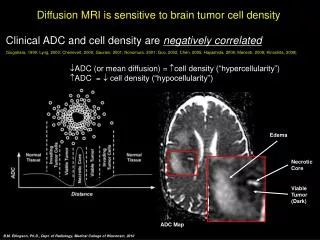
International Journal of Trend in Scientific Research and Development (IJTSRD) Volume 3 Issue 5, August 2019 Volume 3 Issue 5, August 2019 Available Online: www.ijtsrd.com e- International Journal of Trend in Scientific Research and Development (IJTSRD) International Journal of Trend in Scientific Research and Development (IJTSRD) -ISSN: 2456 – 6470 Brain Tumor Detection System Moe Moe Aye Moe Moe Aye1, Kyaw Kyaw Lin2 Brain Tumor Detection System for MRI Image or MRI Image 1Department of Information Technology, 1, 2Technological University echnological University, Mandalay, Myanmar f Information Technology, 2Department of Mechatronic Engineering f Mechatronic Engineering, How to cite this paper: Moe Moe Aye | Kyaw Kyaw Lin "Brain Tumor Detection System for MRI Image" Published in International Journal of Trend in Scientific Research and Development (ijtsrd), ISSN: 2456- 6470, Volume-3 | Issue-5, August 2019, pp.2070-2073, https://doi.org/10.31142/ijtsrd27864 Copyright © 2019 by author(s) and International Journal of Trend in Scientific Research and Development Journal. This is an Open Access article distributed under the terms of the Creative Commons Attribution License (CC (http://creativecommons.org/licenses/by /4.0) The brain tumor detection is very hard in beginning st because it cannot find the accurate measurement of tumor. Thus, the brain tumor detection still remains challenging problem due to complex structure of brain than other breast, heart, lung, kidney, bone cancer detections in medical fields. Tumor segmentation from MRI data is an important process but it is time-consuming and difficult task often performed manually by medical experts. Radiologists and other medical experts spend a substantial amount of time segmenting medical images when a large number of MRI brain images are analyzed. Moreover, accurately labeling brain tumor may lead to missing diagnosis. And it is a considerable variation that is observed between doctors. To avoid human based diagnostic error, computer aided diagnosis system is needed. Throughout the few years, different segmentation methods have been used for tumor detection but it is time consuming process and also gives inaccurate result. And most of them fail because of unknown noises, poor image contrast, in homogeneity and weak boundaries that are usual in medical images. In this paper, the Brain Tumor Detection System is designed to detect the brain tumor from MRI brain images accurately in order to enhance the performance of the image segmentation especially in the field of brain segmentation. Implementation and identification of brain tumor are done by segmentation and detecting the brain image from the MRI scans. In the system, pre-processing of the image is the primary step which removes noises and smoothes the input MRI brain image. And, segmentation is carried out using thresholding technique. Then, the brain tumor region is detected from the segmented image using region properties. detected from the segmented image using region properties. ABSTRACT Today, computer aided system is widely used in various fields. Among them, the brain tumor detection is an important task in medical image processing. Early diagnosis of brain tumors plays an important role in improving treatment possibilities and increases the survival rate of the patients. Manual segmentation of brain tumors for cancer diagnosis, from large amount of Magnetic Resonance Imaging (MRI) images generated in c difficult and time consuming task or even generates errors. So, the automatic brain tumor segmentation is needed to segment tumor. The purpose of the thesis is to detect the brain tumor quickly and accurately from the MRI brain image. In the system, the average filter is used to remove noise and make smooth an input MRI image and threshold segmentation is applied to segment tumor region from MRI brain images. Region properties method is used to detect the tumor region exactly. And then, the equation of the tumor region in the system is effectively applied in any shape of the tumor region. KEYWORDS: area of tumor; brain tumor detection; location in brain; MRI image; segmentation; tumor region INTRODUCTION Medical imaging plays a central role in the diagnosis of brain tumors. Brain tumor diagnosis is quite difficult because of diverse shape, size, location and appearance of tumor in brain. Today, computer aided system is widely used in various fields. Among them, the brain tumor detection is an important task in medical image processing. brain tumors plays an important role in improving treatment possibilities and increases the survival rate of the patients. Manual segmentation of brain tumors for cancer diagnosis, from large amount of Magnetic Resonance Imaging (MRI) images generated in clinical routine, is a difficult and time consuming task or even generates errors. So, the automatic brain tumor segmentation is needed to segment tumor. The purpose of the thesis is to detect the brain tumor quickly and accurately from the MRI brain In the system, the average filter is used to remove noise and make smooth an input MRI image and threshold segmentation is applied to segment tumor region from MRI brain images. Region properties method is used to IJTSRD27864 , the equation of the tumor region in the system is effectively applied in any shape of the tumor region. area of tumor; brain tumor detection; location in brain; MRI image; central role in the diagnosis of brain tumors. Brain tumor diagnosis is quite difficult because of diverse shape, size, location and BY 4.0) http://creativecommons.org/licenses/by The brain tumor detection is very hard in beginning stage because it cannot find the accurate measurement of tumor. Thus, the brain tumor detection still remains challenging problem due to complex structure of brain than other breast, heart, lung, kidney, bone cancer detections in medical fields. tation from MRI data is an important process consuming and difficult task often performed manually by medical experts. Radiologists and other medical experts spend a substantial amount of time segmenting Finally, the area and location of the brain tumor are calculated. The purpose of the system is to assist doctors and radiologists who examine and determine the symptoms of brain tumor in biomedical field by using image processing techniques. Therefore, once it gets identified brain tumor, it gives to start the proper treatment Finally, the area and location of the brain tumor are calculated. The purpose of the sys radiologists who examine and determine the symptoms of brain tumor in biomedical field by using image processing techniques. Therefore, once it gets identified brain tumor, it gives to start the proper treatment and it may be curable. RELATED WORK This session presents the related work for the detection of brain tumor in digital image processing. K., Surabhi [1] have proposed an efficient brain tumor detection system based on the segmentation technique for MRI brain images, which can detect tumor and trace the position of tumor in the MRI brain images. To detect the tumor region, segmentation method is used o threshold and it exactly detects the tumor region with better accuracy. In their proposed work, some pre (resizing, binarization and morphological operations) are used for detection of tumor region in MRI brain images. Their study introduces an efficient detection of brain tumor from MRI brain images based on the segmentation of gray matter and white matter. The purpose of this segmentation algorithm is to label tumor objects within the MRI brain image and locate their region tumor is extracted from the MRI brain images and its exact position is also determined to classify the volume of tumor. By experimental analysis of various parameters such as PSNR, MSE and volume or percentage of tumor, they observed that the accuracy of their proposed system is more that 90% and it is better than the previous work. that 90% and it is better than the previous work. MRI brain images This session presents the related work for the detection of brain tumor in digital image processing. are analyzed. Moreover, accurately labeling brain tumor may lead to missing diagnosis. And it is a considerable variation that is observed between doctors. To avoid human based diagnostic error, computer aided diagnosis system is needed. Throughout the few years, different segmentation methods have been used for tumor detection but it is time consuming process and also gives inaccurate result. And most of them fail because of unknown noises, poor image contrast, in ndaries that are usual in medical images. In this paper, the Brain Tumor Detection System is designed to detect the brain tumor from MRI brain images accurately in order to enhance the performance of the image segmentation especially in the field of brain tissue [1] have proposed an efficient brain tumor detection system based on the segmentation technique for MRI brain images, which can detect tumor and trace the position of tumor in the MRI brain images. To detect the tumor region, segmentation method is used on the basis of threshold and it exactly detects the tumor region with better accuracy. In their proposed work, some pre-processing steps (resizing, binarization and morphological operations) are used for detection of tumor region in MRI brain images. study introduces an efficient detection of brain tumor from MRI brain images based on the segmentation of gray matter and white matter. The purpose of this segmentation algorithm is to label tumor objects within the MRI brain image and locate their region based on the white matter. The tumor is extracted from the MRI brain images and its exact position is also determined to classify the volume of tumor. By experimental analysis of various parameters such as PSNR, MSE and volume or percentage of tumor, they have observed that the accuracy of their proposed system is more Implementation and identification of brain tumor are done by segmentation and detecting the brain image from the MRI processing of the image is the primary step which removes noises and smoothes the input brain image. And, segmentation is carried out using thresholding technique. Then, the brain tumor region is @ IJTSRD | Unique Paper ID – IJTSRD27864 27864 | Volume – 3 | Issue – 5 | July - August 2019 August 2019 Page 2070
International Journal of Trend in Scientific Research and Development (IJTSRD) @ www.ijtsrd.com eISSN: 2456-6470 Devkota, et al. [2] have proposed a computer aided detection approach to diagnose brain tumor in its early stage using Mathematical Morphological Reconstruction (MMR). Image is pre-processed to remove noise and artifacts and then segmented to find regions of interest with probable tumor. A large number of textural and statistical features are extracted from the segmented image to classify whether the brain tumor in the image is benign or malignant. Their experimental results show that the segmented images have a high accuracy while substantially reducing the computation time. Their study shows that the proposed solution can be used to diagnose brain tumor in patients with a high success rate. A., Meena, and K., Raja proposed an approach of Spatial Fuzzy C-Means (PETSFCM) clustering algorithm on Positron Emission Tomography (PET) scan image datasets. The algorithm is joining the spatial neighborhood information with classical Fuzzy C-Means (FCM) and updating the objective function of each cluster. Spatial relationship of neighboring pixel is an aid of image segmentation. These neighboring pixels are highly renovated the same feature data. In spatial domain, the member-ships of the neighbor centered are specified to obtain the cluster distribution statistics. They calculated the weighting function based on these statistics and applied into the member-ship function. Their algorithm is tested on data collection of patients with Alzheimer’s disease. They did not calculate objective based quality assessment that could analyze images and did not report their quality without human involvement. METHODOLOGY This session expresses the background theory used in the brain tumor detection system. Average filter, image segmentation, region properties and calculation of tumor area in MRI brain images are explained in the following sections. A.Average Filter The default filter type is the average or mean filter in the general class. The average filter is low pass filter. The output cell value calculated by filter is the simple average (arithmetic mean) of the cells in the filter window. The averaging performed by the average filter removes some of the higher frequency features, while allowing the low- frequency features to pass through the filter unchanged (thus the term “low pass” filter). This has the effect of smoothing the raster image, emphasizing its larger-scale brightness trends. Whether the smoothing produced by the low pass filter is beneficial or not depends on the characteristics of the input image. Average filter is used to smooth the image and remove noises such as Gaussian noise. It reduces the amount of intensity variation between one pixel and the next. Average filter is an estimation of average from neighbor pixels within the kernel. The filtering equation can be expressed as (1). g(x,y) = 1/M∑(x,y)∈S f(x,y) B.Image Segmentation Thresholding is one of the commonly used methods for image segmentation [5]. Image threshold is a simple, effective and way of partitioning an image into a foreground and background. This image analysis technique is a type of image segmentation that isolates objects by converting grayscale images into binary images. Regions of an image are separated out corresponding to objects which want to analyze. Each pixel is labeled as belonging to one of (typically) two classes. At present, threshold-based methods are classified into global, local and adaptive thresholdings. In this study, global threshold based segmentation method is used to segment brain tumor in MRI brain image. Global (single) thresholding method is used when the intensity distribution between the objects of foreground and background are very distinct. When the differences between foreground and background objects are very distinct, a single value of threshold can simply be used to differentiate both objects apart [6]. Global methods find a single threshold value T for the whole document. Then each pixel is assigned to page foreground or background based on its gray value comparing with the threshold value. Global methods are very fast and they give good results for typical scanned documents. For many years, the binarization of a grayscale document was based on the global thresholding statistical algorithms [7]. Some most common used global thresholding methods are Otsu method, entropy based thresholding, etc. Otsu’s algorithm is a popular global thresholding technique. Moreover, there are many popular thresholding techniques such as Kittler and Illingworth, Kapur, Tasi, Huang, Yen and et al [8]. The condition for selecting the T is given as (2). g(x,y) = ?1 if f(x,y) > T 0 otherwise (2) where g(x, y) is the thresholded image, f (x, y) is the input image, and T is the threshold value. Equation (2) has no indication on selecting the threshold value T. The threshold T separates the object from the dark background. Any point (x,y) for which f(x, y) >T is called an object point. Pixels are labeled 1 corresponds to object whereas pixels labeled 0 corresponds to the background [8]. Global thresholding is popular due to simplicity and easy implementation [5][9]. C.Region Properties Image regions are also called objects, connected components, or blobs, can be contiguous or discontiguous. It returns measurements for the set of properties specified by properties for 8-connected component (object) in the binary image, black and white. A region in an image can have properties, such as Area, BoundingBox, ConvexArea, Centroid, Eccentricity, ConvexHull, EulerNumber, Extent, Extrema, FilledImage, MajorAxisLength, Orientation, PixelList, PixelIdxList, Solidity, SubarrayIdx, Perimeter, ConvexImage. Measurement values are returned as an array of structure or a table. Region properties can be a comma-separated list of strings, a cell array containing strings, the single string 'all', or the string 'basic'. If properties are the string 'all', then all the preceding measurements are computed. If properties are not specified or are the string 'basic', then these measurements are computed: 'Area', 'Centroid', and 'BoundingBox' [10]. EquivDiameter, FilledArea, MinorAxisLength, Image, (1) where S is the neighborhood of pixel (x,y), M is the number of pixels in neighborhood S, and g(x,y) is the filtered image. Sub-images are summed up and then multiplied by 1/M [4]. @ IJTSRD | Unique Paper ID – IJTSRD27864 | Volume – 3 | Issue – 5 | July - August 2019 Page 2071
International Journal of Trend in Scientific Research and Development (IJTSRD) @ www.ijtsrd.com eISSN: 2456-6470 D.Area Calculation of Tumor Region The tumor area is calculated using the binarization method. That is the image having only two values either black or white (0 or 1) [11]. 256x256 jpeg image is the maximum image size. The binary image can be represented as a summation of total number of white and black pixels shown in (3). The number of white pixels is calculated by (4). Image I=∑ ∑ [f(0)+f(1)] H=0 W=0 where f(0) is black pixel (digit 0) and f(1) is white pixel (digit 1). P = ∑ ∑ [f(0)] ??? ??? At first, MRI image of the brain is accepted as input image of the system. In pre-processing step, the input MRI brain image is converted into grayscale image. When MRI/CT images are viewed, they look like black and white but they contain some primary colors (RGB). Therefore, grayscale conversion is needed in this step. This result is put to the filtering step using average filter for smoothing the input image. Several different filters can be used for filtering. But, MRI image does not contain a lot of noise. So, the average filter is used in this step. After using the average filter, the smooth image is obtained. This smooth image is used to operate the next step of the system quickly. After removing the noise from the input brain image, this image is loaded into the segmentation step. In this step, global thresholding is used and the threshold point 0.6 is selected as standard threshold point for this segmentation step. This threshold point is very effective for all MRI brain images. If a suitable threshold point is not chosen, the detection of the brain tumor region can be difficult to find the accurate tumor region. Therefore, an appropriate threshold point is chosen for the system. The segmented images can have pixels above the standard threshold point. The image from the segmentation process is labeled to use region properties. Many region properties of the image may be used to detect the tumor region, but only two region properties are used in the system such as ‘Area’ and ‘Solidity’. Solidity gives the proportion of the pixels in the convex hull that are also in the region. So, solidity is what fraction of the actual area region is. For any convex object it’s 1. An asterisk might have a solidity of around 0.5, while a thin “L” shape (or “T” or “E”, etc.) would have a very low solidity. Moreover, solidity also helps to find the normal brain which is no tumor in the tumor detection stage. And then area of the region properties is used to find the member of the result from the defined solidity value. After these steps are finished, the brain tumor region is obtained quickly and accurately. In this step, the output from the detection process is used to calculate the area of the tumor region in the brain slice. The size of images, 256 × 256, are used as the maximum image size for the system so that the dimensions of the MRI brain images are equal size. Therefore, the calculation of the tumor region is more efficient to analyze for medical fields. This region properties method generates automatically tumor area in pixels. So the white pixels of tumor region from the detected brain image are without needed to count. Finally, the location of the tumor region can be determined to know position whether tumor is at left hemisphere, right hemisphere or near the center of the brain. The column pixels or x-dimension of the first input image from the system is divided by 2. The minimum value and the maximum value are calculated to compare with the center value of image which is divided result of input image. If the tumor is located in the left part of the brain, the tumor is actually located in the right hemisphere of the brain or inversely. If the minimum value is greater than the center value, the tumor is located in the left hemisphere of the brain. If the maximum value is less than the center value, the tumor is located in the right hemisphere of the brain. Otherwise, the tumor is located near the center of the brain. 255 255 (3) (4) ??? ??? , where P is number of white pixel and 1 Pixel is equal to 0.264 mm. The area calculation formula is shown in (5). Size_of_tumor_is, S=[(√A)*0.264]mm2 DESIGN OF THE SYSTEM The typical brain tumor detection generally consists of five basic steps: image pre-processing, tumor segmentation, tumor detection, calculation of the brain tumor area and determination of the brain tumor location. The flow chart of the system is shown in Fig. 1. (5) Figure1. Flow Chart of the Proposed System @ IJTSRD | Unique Paper ID – IJTSRD27864 | Volume – 3 | Issue – 5 | July - August 2019 Page 2072
International Journal of Trend in Scientific Research and Development (IJTSRD) International Journal of Trend in Scientific Research and Development (IJTSRD) @ www.ijtsrd.com www.ijtsrd.com eISSN: 2456-6470 IMPLEMENTATION RESULTS There are forty-seven MRI brain images collected to detect brain tumor. Thirty-six brain tumor images and eleven normal brain images are tested. The step by step results of the brain tumor detection system are shown in Fig. 2. cancer the patient suffers and to take necessary and appropriate treatment steps. cancer the patient suffers and to take necessary and appropriate treatment steps. REFERENCES [1]K. Surabhi, “An Efficient Brain Tumor Detection System Based on Segmentation Technique for MRI Brain Images,” International Journal of Advanced Research in Computer Science, Volume 8, No. 7, pp. 1131 – August 2017. seven MRI brain images collected to detect six brain tumor images and eleven normal brain images are tested. The step by step results of the brain tumor detection system are shown in Fig. 2. K. Surabhi, “An Efficient Brain Tumor Detection System Based on Segmentation Technique for MRI Brain Images,” International Journal of Advanced Research in Computer Science, Volume 8, No. 7, pp. 1131-1136, July [2]Devkota, et al., “Image Segmentation for Early Stage Brain Tumor Detection Morphological Reconstruction”, Procedia Computer Science, vol. 125. Pp. 115 Science, vol. 125. Pp. 115-123, 2018. mage Segmentation for Early Stage Detection Morphological Reconstruction”, Procedia Computer Brain Tumor using using Mathematical Mathematical [3]Meena, and K. Raja, “Spatial Fuzzy C Segmentation International Journal of Computer Science and Engineering (IJCSE), 2013. Engineering (IJCSE), 2013. Meena, and K. Raja, “Spatial Fuzzy C-means PET Image Segmentation International Journal of Computer Science and of of Neurodegenerative Neurodegenerative Disorde Disorder”, Figure2. Results of the Brain Tumor Detection rain Tumor Detection [4]Gonzalez, et al.:“Digital Image Processing”, Second Edition, Prentice Hall 2002 Edition, Prentice Hall 2002 Gonzalez, et al.:“Digital Image Processing”, Second If the tumor is detected, the area and location of the brain tumor region are calculated and displayed. If not, the result will be displayed as Fig. 3. If the tumor is detected, the area and location of the brain tumor region are calculated and displayed. If not, the result [5]S. A. Salem, N. V. Kalyankar, and S. D. Khamitkar, “Image Segmentation by using Thershod Techniques Journal of Computing, vol2, pp 83 Journal of Computing, vol2, pp 83-86, 2010 S. A. Salem, N. V. Kalyankar, and S. D. Khamitkar, “Image Segmentation by using Thershod Techniques”, [6]N. N. Segmentation by Using Thresholding Techniques for Medical Images”, Computer Science & Engineering: An International Journal (CSEIJ), Vol.6, No.1, February Senthilkumaran, Segmentation by Using Thresholding Techniques for Medical Images”, Computer Science & Engineering: An International Journal (CSEIJ), Vol.6, No.1, February 2016 Senthilkumaran, and and S. S. Vaithegi, Vaithegi, “Image “Image [7]G. S. Evelin, Y. V. S. Lakshmi, and G. J. Wiselin, “MRI Brain Image Segmentation based on Thresholding”, International Journal of Advanced Computer Research, Volume-3 Number-1, Issue 1, Issue-8, March-2013 velin, Y. V. S. Lakshmi, and G. J. Wiselin, “MRI Brain Image Segmentation based on Thresholding”, International Journal of Advanced Computer Research, [8]G. K. Priyanka, and N. H. Sushilkumar, “A Review of Image Thresholding Techniques”, International Journal of Advanced Research in Computer Science and Software Engineering, Volume 5, Issue 6, June 2015 Software Engineering, Volume 5, Issue 6, June 2015 G. K. Priyanka, and N. H. Sushilkumar, “A Review of Thresholding Techniques”, International Journal of Advanced Research in Computer Science and Figure3. Result for Normal Brain 3. Result for Normal Brain CONCLUSION The brain tumor detection system for MRI images has been presented in this paper. The calculation of tumor area plays a vital role in assisting the treatment planning. The stage of tumor is based on the area of tumor. In the proposed system, till the small size of brain tumor, 3 mm2, can we found that if the area of the brain tumor is greater than 6 mm2, it will be critical position. The segmentation and detection methods are low cost as it can be implemented in general computer. Although the computer aided techniques are complex to be implemented but not as tedious, laborious, and time consuming as manual methods. Therefore, the system will help doctors to take or analyze in which stage of system will help doctors to take or analyze in which stage of [9]S. Abutaleb, “Automatic Thresholding of Gray Pictures using Two Dimensional Entropy”, Computer Vision, Grapics, and Image processing, vol.47, pp. 22 32, 1989 S. Abutaleb, “Automatic Thresholding of Gray -Level Pictures using Two Dimensional Entropy”, Computer apics, and Image processing, vol.47, pp. 22- for MRI images has been presented in this paper. The calculation of tumor area plays a vital role in assisting the treatment planning. The stage of tumor is based on the area of tumor. In the proposed system, till the small size of brain tumor, 3 mm2, can be detected and we found that if the area of the brain tumor is greater than 6 mm2, it will be critical position. The segmentation and detection methods are low cost as it can be implemented in general computer. Although the computer aided techniques omplex to be implemented but not as tedious, laborious, and time consuming as manual methods. Therefore, the [10]Anonymous, MathWorks, Inc., 2018 Anonymous, “Image “Image Region Region Properties”. Properties”. The The [11]J. Selvakumar, A. Lakshmi, and T. Arivoli, “Brain Tumor Segmentation and Its Area Calculation in Brain MR Images using K-Mean Clustering an Algorithm”, IEEE-International Advances In Engineering, Science And Management (ICAESM -2012) March 30, 31, 2012 2012) March 30, 31, 2012 J. Selvakumar, A. Lakshmi, and T. Arivoli, “Brain Tumor Segmentation and Its Area Calculation in Brain MR Mean Clustering and Fuzzy C-Mean International Advances In Engineering, Science And Management Conference Conference On On @ IJTSRD | Unique Paper ID – IJTSRD27864 27864 | Volume – 3 | Issue – 5 | July - August 2019 August 2019 Page 2073