Download
1 / 24
290 likes | 506 Views
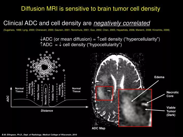
Diffusion MRI is sensitive to brain tumor cell density. Clinical ADC and cell density are negatively correlated (Sugahara, 1999; Lyng, 2000; Chenevert, 2000; Gaurain, 2001; Nonomura, 2001; Guo, 2002; Chen, 2005; Hayashida, 2006; Manenti, 2008; Kinoshita, 2008)

E N D
Diffusion MRI is sensitive to brain tumor cell density • Clinical ADC and cell density are negatively correlated • (Sugahara, 1999; Lyng, 2000; Chenevert, 2000; Gaurain, 2001; Nonomura, 2001; Guo, 2002; Chen, 2005; Hayashida, 2006; Manenti, 2008; Kinoshita, 2008) • ADC (or mean diffusion) = cell density (“hypercellularity”) • ADC = cell density (“hypocellularity”) Edema Necrotic Core Viable Tumor (Dark) ADC Map B.M. Ellingson, Ph.D., Dept. of Radiology, Medical College of Wisconsin, 2010
Diffusion MRI is sensitive to brain tumor cell density • Tests at our laboratory have confirmed this relationship • 17 glioma patients (WHO II-IV) underwent diagnostic stereotactic biopsy • Biopsy sites were spatially matched to pre-operative ADC maps R2 = 0.7933; P < 0.001 From: Ellingson, et al. JMRI 2009, In Press SNO/CNS 2009 B.M. Ellingson, Ph.D., Dept. of Radiology, Medical College of Wisconsin, 2010
The Functional Diffusion Map (fDM)(Moffat, 2005; 2006; Hamstra, 2005; 2008) From: Ellingson, JMRI, 2009, In Press B.M. Ellingson, Ph.D., Dept. of Radiology, Medical College of Wisconsin, 2010
SNO/CNS 2009 B.M. Ellingson, Ph.D., Dept. of Radiology, Medical College of Wisconsin, 2009 B.M. Ellingson, Ph.D., Dept. of Radiology, Medical College of Wisconsin, 2010
SNO/CNS 2009 B.M. Ellingson, Ph.D., Dept. of Radiology, Medical College of Wisconsin, 2009 B.M. Ellingson, Ph.D., Dept. of Radiology, Medical College of Wisconsin, 2010 From: Ellingson et al. J Neurooncol, 2009, In Press
Early Detection of Brain Tumor Growth T1+C Contrast-Enhancement (white) FLAIR Hypercellular Regions (Blue) fDMs B.M. Ellingson, Ph.D., Dept. of Radiology, Medical College of Wisconsin, 2010
fDMs in Brain Tumor Progression 3 mo. 6 mo. 9 mo. (Onset of symptoms) T1+C FLAIR fDM B.M. Ellingson, Ph.D., Dept. of Radiology, Medical College of Wisconsin, 2010
fDMs in Progressive Disease (PD) Hypercellularity Hypercellularity Hypercellularity SNO/CNS 2009 B.M. Ellingson, Ph.D., Dept. of Radiology, Medical College of Wisconsin, 2010
fDM Results in Stable Disease (SD) Treatment: Radiation + Temozolomide SNO/CNS 2009 B.M. Ellingson, Ph.D., Dept. of Radiology, Medical College of Wisconsin, 2010
fDM Results in Responding Disease (RD) Treatment: Radiation + Temozolomide SNO/CNS 2009 B.M. Ellingson, Ph.D., Dept. of Radiology, Medical College of Wisconsin, 2010
fDM Results in Stable/Responding Disease (SD/RD) Hypocellularity Hypocellularity Hypocellularity SNO/CNS 2009 B.M. Ellingson, Ph.D., Dept. of Radiology, Medical College of Wisconsin, 2010
fDMs are an early biomarker for cytotoxic and new anti-angiogenic treatments B.M. Ellingson, Ph.D., Dept. of Radiology, Medical College of Wisconsin, 2010 Ellingson BM, J Neurooncol, Under Prep
Results • “fDM Responders” have significantly longer TTP after standard Tx • fDMs are a better predictor than tumor grade SNO/CNS 2009 B.M. Ellingson, Ph.D., Dept. of Radiology, Medical College of Wisconsin, 2010
Results • “fDM Responders” have significantly longer survival on bevacizumab • fDMs are better predictors than grade, age, or mono/combined therapy SNO/CNS 2009 B.M. Ellingson, Ph.D., Dept. of Radiology, Medical College of Wisconsin, 2010
Graded fDMs Allow Visualization of Growing Tumor 1 Mo. 2 Mo. 3 Mo. 4 Mo. + Hypocellular + Hypercellular B.M. Ellingson, Ph.D., Dept. of Radiology, Medical College of Wisconsin, 2010
Graded fDMs Allow Visualization of Growing Tumor 5 Mo. 7 Mo. 3 Mo. + Hypocellular + Hypercellular B.M. Ellingson, Ph.D., Dept. of Radiology, Medical College of Wisconsin, 2010
Graded fDMs Allow Visualization of Growing Tumor 4 Mo. 5 Mo. 2 Mo. + Hypocellular + Hypercellular B.M. Ellingson, Ph.D., Dept. of Radiology, Medical College of Wisconsin, 2010
Graded fDMs in Demyelination 3 Mo. 5 Mo. 7 Mo. 9 Mo. Hypercellular Hypocellular Macrophages & Inflammatory Cells Demyelination Biopsy Diagnosis = Demyelination (Multiple Sclerosis) B.M. Ellingson, Ph.D., Dept. of Radiology, Medical College of Wisconsin, 2010
Graded fDMs: Radiation Necrosis vs. Tumor T1+C FLAIR Hypercellular Graded fDM Hypocellular B.M. Ellingson, Ph.D., Dept. of Radiology, Medical College of Wisconsin, 2010
Graded fDMs: Radiation Induced Changes 2 Mo. 3 Mo. 5 Mo. 8 Mo. 14 Mo. 18 Mo. 21 Mo. 23 Mo. Hypercellular Hypocellular B.M. Ellingson, Ph.D., Dept. of Radiology, Medical College of Wisconsin, 2010
Graded fDMs: Radiation Induced Changes 2 Mo. 4 Mo. 6 Mo. 14 Mo. 10 Mo. 18 Mo. Hypercellular Hypocellular B.M. Ellingson, Ph.D., Dept. of Radiology, Medical College of Wisconsin, 2010
Graded fDMs Improve Tumor Localization/Grading in Stereotactic Needle Biopsy B.M. Ellingson, Ph.D., Dept. of Radiology, Medical College of Wisconsin, 2010
Graded fDMs Improve Tumor Localizationand Grading in Stereotactic Needle Biopsy Biopsy (WHO II) 2 Lesions Hypercellular Hypocellular B.M. Ellingson, Ph.D., Dept. of Radiology, Medical College of Wisconsin, 2010
Graded fDMs Improve Tumor Localization for Resection Resection Cavity Hypercellular Only 10% of Hypercellular regions were removed! Hypocellular B.M. Ellingson, Ph.D., Dept. of Radiology, Medical College of Wisconsin, 2010