Download
1 / 5
50 likes | 58 Views
Brain tumor segmentation is one of the critical tasks in the medical image processing. Some early diagnosis of brain tumor helps in improving the treatment and also increases the survival rate of the patients. The manual segmentation for cancer diagnosis of brain tumor and generation of MRI images in clinical routine is difficult and time consuming. The aim of this research paper is to review of MRI based brain tumor segmentation methods for the treatment of cancer like diseases. The magnetic resonance imaging used for detection of tumor and diagnosis of tissue abnormalities. The computerized medical image segmentation helps the doctors in treatment in a simple way with fast decision making. The brain tumor segmentation assessed by computer based surgery, tumor growth, developing tumor growth models and treatment responses. This research focuses on the causes of brain tumor, brain tumor segmentation and its classification, MRI scanning process and different segmentation methodologies. Ishu Rana | Gargi Kalia | Preeti Sondhi "MRI Image Segmentation by Using DWT for Detection of Brain Tumor" Published in International Journal of Trend in Scientific Research and Development (ijtsrd), ISSN: 2456-6470, Volume-3 | Issue-4 , June 2019, URL: https://www.ijtsrd.com/papers/ijtsrd25116.pdf Paper URL: https://www.ijtsrd.com/computer-science/bioinformatics/25116/mri-image-segmentation-by-using-dwt-for-detection-of-brain-tumor/ishu-rana<br>

E N D
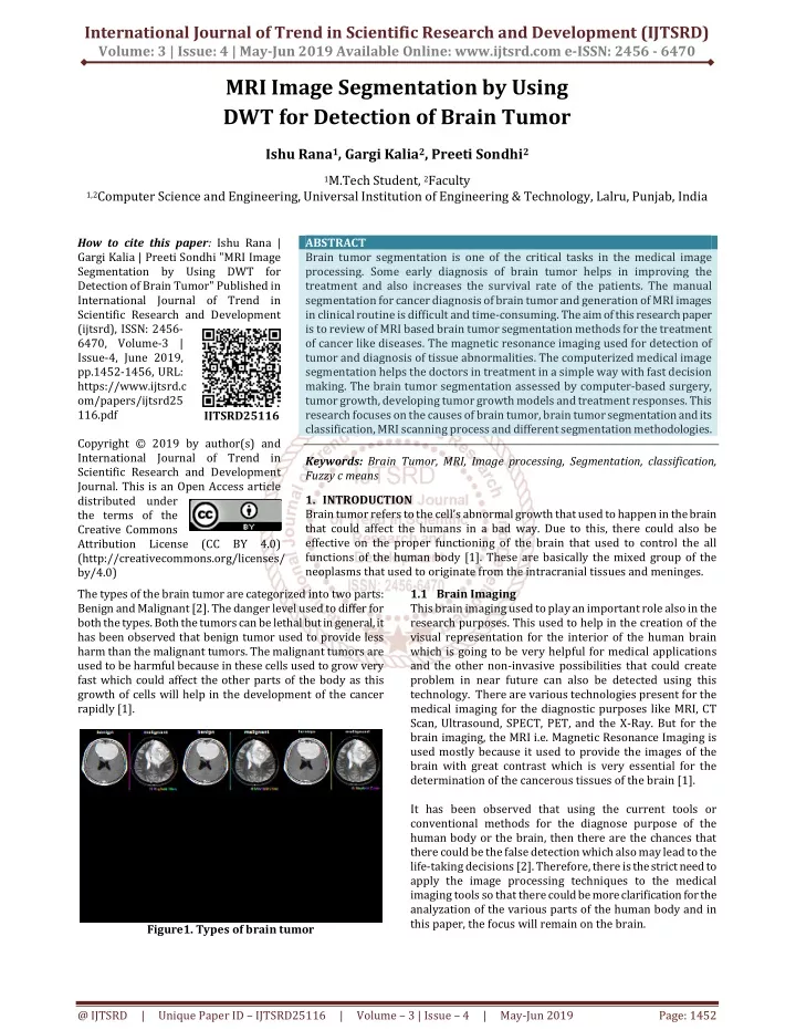
International Journal of Trend in Scientific Research and Development (IJTSRD) Volume: 3 | Issue: 4 | May-Jun 2019 Available Online: www.ijtsrd.com e-ISSN: 2456 - 6470 MRI Image Segmentation by Using DWT for Detection of Brain Tumor Ishu Rana1, Gargi Kalia2, Preeti Sondhi2 1M.Tech Student, 2Faculty 1,2Computer Science and Engineering, Universal Institution of Engineering & Technology, Lalru, Punjab, India How to cite this paper: Ishu Rana | Gargi Kalia | Preeti Sondhi "MRI Image Segmentation by Using DWT for Detection of Brain Tumor" Published in International Journal of Trend in Scientific Research and Development (ijtsrd), ISSN: 2456- 6470, Volume-3 | Issue-4, June 2019, pp.1452-1456, URL: https://www.ijtsrd.c om/papers/ijtsrd25 116.pdf Copyright © 2019 by author(s) and International Journal of Trend in Scientific Research and Development Journal. This is an Open Access article distributed under the terms of the Creative Commons Attribution License (CC BY 4.0) (http://creativecommons.org/licenses/ by/4.0) The types of the brain tumor are categorized into two parts: Benign and Malignant [2]. The danger level used to differ for both the types. Both the tumors can be lethal but in general, it has been observed that benign tumor used to provide less harm than the malignant tumors. The malignant tumors are used to be harmful because in these cells used to grow very fast which could affect the other parts of the body as this growth of cells will help in the development of the cancer rapidly [1]. ABSTRACT Brain tumor segmentation is one of the critical tasks in the medical image processing. Some early diagnosis of brain tumor helps in improving the treatment and also increases the survival rate of the patients. The manual segmentation for cancer diagnosis of brain tumor and generation of MRI images in clinical routine is difficult and time-consuming. The aim of this research paper is to review of MRI based brain tumor segmentation methods for the treatment of cancer like diseases. The magnetic resonance imaging used for detection of tumor and diagnosis of tissue abnormalities. The computerized medical image segmentation helps the doctors in treatment in a simple way with fast decision making. The brain tumor segmentation assessed by computer-based surgery, tumor growth, developing tumor growth models and treatment responses. This research focuses on the causes of brain tumor, brain tumor segmentation and its classification, MRI scanning process and different segmentation methodologies. Keywords: Brain Tumor, MRI, Image processing, Segmentation, classification, Fuzzy c means 1.INTRODUCTION Brain tumor refers to the cell’s abnormal growth that used to happen in the brain that could affect the humans in a bad way. Due to this, there could also be effective on the proper functioning of the brain that used to control the all functions of the human body [1]. These are basically the mixed group of the neoplasms that used to originate from the intracranial tissues and meninges. 1.1 Brain Imaging This brain imaging used to play an important role also in the research purposes. This used to help in the creation of the visual representation for the interior of the human brain which is going to be very helpful for medical applications and the other non-invasive possibilities that could create problem in near future can also be detected using this technology. There are various technologies present for the medical imaging for the diagnostic purposes like MRI, CT Scan, Ultrasound, SPECT, PET, and the X-Ray. But for the brain imaging, the MRI i.e. Magnetic Resonance Imaging is used mostly because it used to provide the images of the brain with great contrast which is very essential for the determination of the cancerous tissues of the brain [1]. It has been observed that using the current tools or conventional methods for the diagnose purpose of the human body or the brain, then there are the chances that there could be the false detection which also may lead to the life-taking decisions [2]. Therefore, there is the strict need to apply the image processing techniques to the medical imaging tools so that there could be more clarification for the analyzation of the various parts of the human body and in this paper, the focus will remain on the brain. IJTSRD25116 Figure1. Types of brain tumor @ IJTSRD | Unique Paper ID – IJTSRD25116 | Volume – 3 | Issue – 4 | May-Jun 2019 Page: 1452
International Journal of Trend in Scientific Research and Development (IJTSRD) @ www.ijtsrd.com eISSN: 2456-6470 segmentation and classification from brain computed tomography image is time-consuming [2]. A. Hebli and S. Gupta (2016) presented a survey of detection of brain tumor by using the image processing. Brain tumor affecting several people including old age and early age people. It is the abnormal growth of cell in the brain that limits the function of the brain. With the help of image processing and the advancement of machine learning, a brain tumor can easily be detected early. This paper discussed image processing in detecting brain tumor and determine technologies that help in predict brain tumor [3]. S. Dhanalakshmi and T.Ravichandran (2012) presents a new method for the segmentation of image which is a form of signal processing. This paper demonstrated image segmentation algorithms which are based on the bottleneck method. the first algorithm which is used in this paper is the split-and-merge algorithm in which an image is segmented in various regions and intensity histogram bins. Another algorithm is histogram clustering algorithm in which input variable presents histogram bins. The registration based algorithm used for registered multimodal images [4]. A. Barbhuiya and K. Hemachandran (2018) presented a hybrid image segmentation model using Wavelet KM, FCM, KM, and Wavelet FCM techniques. This paper compared Wavelet FCM clustering with FCM clustering techniques and conventional KM clustering techniques that are used for image segmentation. These algorithms are tested on various images. The proposed methods in this paper are analyzed with the help of a discrete wavelet transform for image features and enhancing digital images. The result indicated that segmentation by using various images clusters wavelet FCM and wavelet KM performs better than traditional FCM and KM clustering algorithms in terms of segmentation accuracy, sensitivity analysis, CPU execution time, and PSNR (Peak Signal to Noise Ratio) [5]. M. Eido and H. Massoud (2016) described MRI image segmentation with the help of FCM and Wavelet transform algorithm. Image segmentation is an important part of image processing. It is a difficult task to segment magnetic resonance imaging images as they have no linear techniques. It is suitable for biomedical research, clinical diagnosis, etc. This paper focuses on segmenting MRI brain images with the help of Fuzzy c-means (FCM) and Stationary Wavelet Transform (SWT) as a clustering technique [6]. I. Despotovic, B. Goossens, and W. Philips (2015) presented the challenges, applications, and methods of MRI segmentation of the human brain. The image segmentation is known as an important task in the analysis of the medical image in clinical applications. Image segmentation is used for measuring as well as visualizing the anatomical structure in brain MRI analysis for analyzing the changes in the brain for surgical planning, delineating pathological regions and image-guided interventions. This paper reviewed the various methods of brain MRI segmentation. It also describes the differences between these methods. The various MRI pre- processing steps were also explained including bias field correction, image registration, and removal of non-brain tissue [7]. S. Shirly and K. Ramesh (2019) reviewed 2D and 3D MRI image segmentation techniques. The magnetic resonance Figure2. Flowchart for Image Processing of Brain Tumor [1] It has been found that with the help of the image processing techniques, there could be easily detection of the tumors of the brain. The steps that used to be followed in the image processing techniques for the brain tumors are mentioned in the above diagram. In the medical image processing, analysis and extraction of brain tumor is a challenging task because the image of the brain is very complicated. Segmentation has a critical role in the medical image processing. One of the most useful medical diagnostic tools which are used for diagnosis of brain and some other medical images is MRI (Magnetic Resonance Imaging). This research paper mainly focuses on working on various segmentation methods that are used for the detection of the tumor. 2. Literature Review P. Gamage (2017) determined a brain tumor with the help of various image processing techniques. The objectives of medical image processing are to determine efficient and correct information about using images with minimum errors. MRI is used to get the images of cancerous tissue of the human body due to its better quality and high resolution as compared to some other imaging technologies. To take the image of a brain tumor by MRI image is difficult due to the complexity of the brain. MRI images used for processed and to segment the brain tumor. Tumour can be segmented by using techniques of image segmentation. The process of determining brain tumor by using MRI images can be divided into four categories such as image classification, pre- processing, feature extraction and image segmentation [1]. S. Josephine (2018) demonstrated brain tumor MRI image as detection and segmentation with the help of a genetic algorithm. The algorithm used in this research is based on the symmetry character of the brain image. The main objective is to detect the edge and position of tumors automatically. The experiments were carried out by using real pictures. The proposed algorithm is faster and efficient to detect the tumor region from MRI brain image. There are various techniques that are formulated for detecting the brain tumor. Tumour @ IJTSRD | Unique Paper ID – IJTSRD25116 | Volume – 3 | Issue – 4 | May-Jun 2019 Page: 1453
International Journal of Trend in Scientific Research and Development (IJTSRD) @ www.ijtsrd.com eISSN: 2456-6470 imaging is mainly used for human organs for early diagnosis of abnormalities. The automatic computer-aided medical image segmentation is mostly used in the medical diagnostics because of the technical advancements in the image processing techniques. Basically, image segmentation is image processing techniques which are used for searching, extracting the image features and mining the medical image records for accurate and better medical diagnostics. Some commonly sued segmentation techniques are clustering based image segmentation, threshold-based image segmentation, region-based image segmentation, edge based image segmentation, artificial neural network based image segmentation, and atlas-based image segmentation. This paper also described the advantages and limitations of different segmentation techniques [10]. 3. Proposed Work The extraction and the analysis of images of tumor in the human brain is one of the most challenging tasks in the Medical Image processing. It happens because the image of the brain is very complicated and very difficult to understand. Segmentation plays the most critical role in the medical image processing. The magnetic resonance imaging is a useful medical diagnostic tool which is used for diagnosis of the brain as well as for some other medical images. This research mainly focuses on working on various segmentation methods that are used for the detection of the tumor. It also presented the comparison of various segmentation techniques on the basis of PSNR [6]. 4. Algorithm The steps of algorithm for proposed work are shown below: A.Read the MRI image. B.Perform Image preprocessing (Filtering and Resize). C.Apply Discrete Wavelet Transform to extract coefficient matrixes (Average, Horizontal, Vertical, and Diagonal). D.Take the Average Matrix. E.Apply K-Mean Clustering Algorithm On Average Matrix to Segment the tumor region from the image. F.Extract Statistical and texture features from the segmented tumor. 5. Methodology There are several methodologies are used in this research paper are described below and flow chart is shown is figure 3. MRI Image: Magnetic Resonance Imaging is a non-invasive technology which produces the detailed anatomical image of three dimensions without using any damaging radiation. It is used to monitor or diagnose the treatment for several conditions within the abdomen, chest, and pelvis. It provides better images of organs as compared to other scanning techniques. It helps in determining the problems in different parts of the body. Figure 3: Flowchart of proposed work Image Preprocessing: Image preprocessing comes with the parts such as read image, resize as well as removing of noise to enhance image quality to perform further operations. It converts image in digital form and also performs some other operations to get an enhanced image and to extract some useful information. Discrete Wavelet Transform: DWT is widely used in image compressing and signal to process. It is known as multi- resolution analysis and it also decomposes images into Scaling functions as well as wavelet coefficients and this will help for image compressing. Image is a two-dimensional matrix consist of pixels, each pixel represents the digital equivalent of image intensity. DWT transforms converts the spatial domain pixels into frequency domain that are represented in multiple sub-bands. Image is in the spatial domain in that adjacent pixel values are highly correlated to each other and they are redundant. So, these redundancies need to be eliminated to compress images. Feature Extraction: It is used in image processing involves reducing the number of resources required to describe a large set of data. It builds derived features intended to be non-redundant and informative. Basically, it is the transformation of the original image to a data set with a decreased number of variable that contains discriminated information. Classifier: Classification is a type of pattern recognition which is used in machine learning. Classification is of two types Supervised (learning where a training set of correctly identified observations is available) and Unsupervised learning procedure is known as clustering (involves grouping data into categories based on some measure of inherent similarity or distance). Clustering: It is a type of Unsupervised learning. It groups a set of objects in a way that objects in the same group are more similar to each other than to those in other groups and these groups known as clusters. @ IJTSRD | Unique Paper ID – IJTSRD25116 | Volume – 3 | Issue – 4 | May-Jun 2019 Page: 1454
International Journal of Trend in Scientific Research and Development (IJTSRD) @ www.ijtsrd.com eISSN: 2456-6470 6. Experimental Results We are performing the image segmentation method on several cases in order to detect the brain tumor to find whether the proposed method is efficient or not to detect the brain tumor in the human being or not. In this section, we have shown the results with images after applying MRI and DWT technique of the image segmentation method in two cases as shown in Table 1. Figure 7 shows the final segmented images. By using the clustering image segmentation, we detect the region in the brain in which the tumor is found. After that, we eliminate another area except for the detected region in order to get the results more clearly. The third image shows the clear image of the detected tumor area in the brain. These results help in better treatment of brain tumor. If the tumor area is detected in the starting stage of cancer, then it helps in treatment at initial stage and treatment of a particular person. Test 1: The input images and the results of segmented results are discussed in this section by using the proposed algorithm and DWT. The below figure 1 shows the loaded image from which the brain tumor has to be detected after performing some operations and analysis of those results. Test 2: Figure 8 shows the second loaded image which is used to detect tumors in the brain after performing techniques of image segmentation. Figure 9 shows the horizontal contents, vertical contents, diagonal contents, and averaged contents which were obtained after performing the DWT technique which decompose the whole image by using wavelet transform. Figure 10 is obtained after performing averaging of coefficients on the images which we get after performing a discrete wavelet transform. The DWT is applied to MRI images which were fused by the proposed method. This technique decomposes image by using wavelet transform. DWT decomposes the image in four different frequency bands namely LL, LH, HL and HH which contains horizontal contents, vertical contents, diagonal contents, and averaged contents. The below figure 5 shows the reverse operation that is taken place in the original image and obtained from combining four decomposed parts by using IDWT. Figure 11 presents the final segmented images. The first image is obtained after detecting the region of the tumor after performing clustering image segmentation. The second image eliminates the other region where the tumor is not found and the third image shows the detected tumor area so that treatment can be done effectively. After performing discrete wavelet transform, averaging of coefficients is applied on obtained images which provide the average image of a brain tumor which is shown in figure 6. Test Image 1 Test Image 2 Figure 4. Loaded Image 1 (Original Image) Figure 8. Loaded Image 2 (Original Image) Figure 5. Equivalent Image Decomposition Using DWT Figure 9. Equivalent Image Decomposition Using DWT Figure 6. Average Image After Performing Averaging of Coefficients Figure 10. Average Image After Performing Averaging of Coefficients @ IJTSRD | Unique Paper ID – IJTSRD25116 | Volume – 3 | Issue – 4 | May-Jun 2019 Page: 1455
International Journal of Trend in Scientific Research and Development (IJTSRD) @ www.ijtsrd.com eISSN: 2456-6470 Figure 7. Final Segmented Images Figure 11. Final Segmented Images Detailed Results The below table shows the statistical features for 4 images such as mean, standard deviation, skewness, kurtosis, energy and entropy. Table2: Statistical features for few images. Images Mean Standard deviation Skewness Kurtosis Energy RMS Entropy Image 1 0.00 0.09 3.21 Image 2 0.01 0.09 2.61 Image 3 0.00 0.09 0.67 Table3: Textural features with Smoothness and Inverse Difference Movements Images Contrast Homogeneity Correlation Smoothness Variance Inverse Difference Movement Image 1 0.40 0.96 0.12 Image 2 0.35 0.95 0.22 Image 3 0.22 0.94 0.16 Conclusion From this research, it is concluded that image segmentation is very important in image processing. It also helps in analyzing the images. Basically, the image segmentation is used to segment the image into various content which is also known as the region of interest. It is concluded the various techniques for image segmentation are efficient in detecting the brain tumor and face recognition. It is also found that image segmentation techniques by using MRI and DWT are effective in detecting the tumor with its exact location which helps in its treatment in an earlier stage. References [1]Praveen Gamage, “Identification of Brain Tumor using Image Processing Techniques”, Research Gate, September 2017. 37.12 26.02 7.90 0.88 0.83 0.77 0.09 0.09 0.09 2.13 2.35 3.49 0.94 0.96 0.93 0.01 0.01 0.01 2.29 0.20 0.33 [5]A. H. M. Jaffar Iqbal Barbhuiya1*, K. Hemachandran2, “Hybrid Image Segmentation Model using KM, FCM, Wavelet KM, and Wavelet FCM Techniques”, Research Gate, Vol.-6, Issue-9, September 2018. [6]M. Eido and H. Massoud, "MRI Image Segmentation using Stationary Wavelet Transform and FCM Algorithm", Higher Institute for Applied Sciences and Technology, pp. 1-10, 2016. [7]Despotović, B. Goossens and W. Philips, "MRI Segmentation of the Human Brain: Challenges, Methods, and Applications", Computational Mathematical Methods in Medicine, vol. 2015, no. 450341, pp. 1-23, 10.1155/2015/450341 and 2015. Available: [8]S. Shirly and K. Ramesh, "Review on 2D and 3D MRI Image Segmentation Techniques", Current Medical Imaging, vol. 15, no. 2, pp. 150 - 160, 2019. [Accessed 3 May 2019]. [2]S. Josephine, “Brain Tumor MRI Image Detection and Segmentation Using Genetic Algorithm”, International Journal of Computer Sciences and Engineering, Volume- 6, Special Issue-2, March 2018. [9]A. Sindhu, S. Meera “A Survey on Detecting Brain Tumor in MRI Images Using Image Processing Techniques”, International Journal of Innovative Research in Computer and Communication Engineering, Vol. 3, Issue 1, January 2015. [3]Amruta Pramod Hebli, Sudha Gupta, “Brain tumor detection using image processing: A SURVEY”, Research Gate, November 2016. [4]S. Dhanalakshmi, Dr. T. Ravichandran, “A New Method for Image Segmentation”, International Journal of Advanced Research in Computer Science and Software Engineering, Volume 2, Issue 9, September 2012. [10]S. Dalmiya, A. Dasgupta, S. K. Datta. “Application of wavelet-based K- means algorithm in mammogram segmentation”. International Journal of Computer Applications, Vol. 52, No. 15, Jan 2012. @ IJTSRD | Unique Paper ID – IJTSRD25116 | Volume – 3 | Issue – 4 | May-Jun 2019 Page: 1456