Download
1 / 4
40 likes | 44 Views
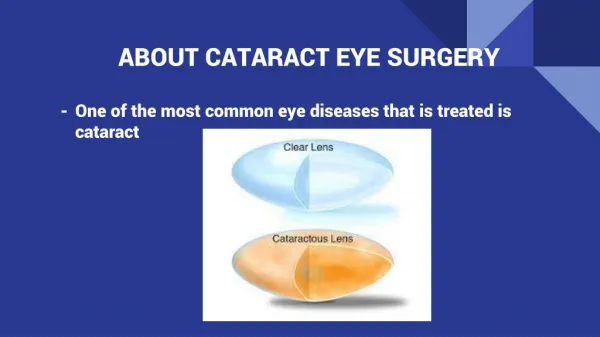
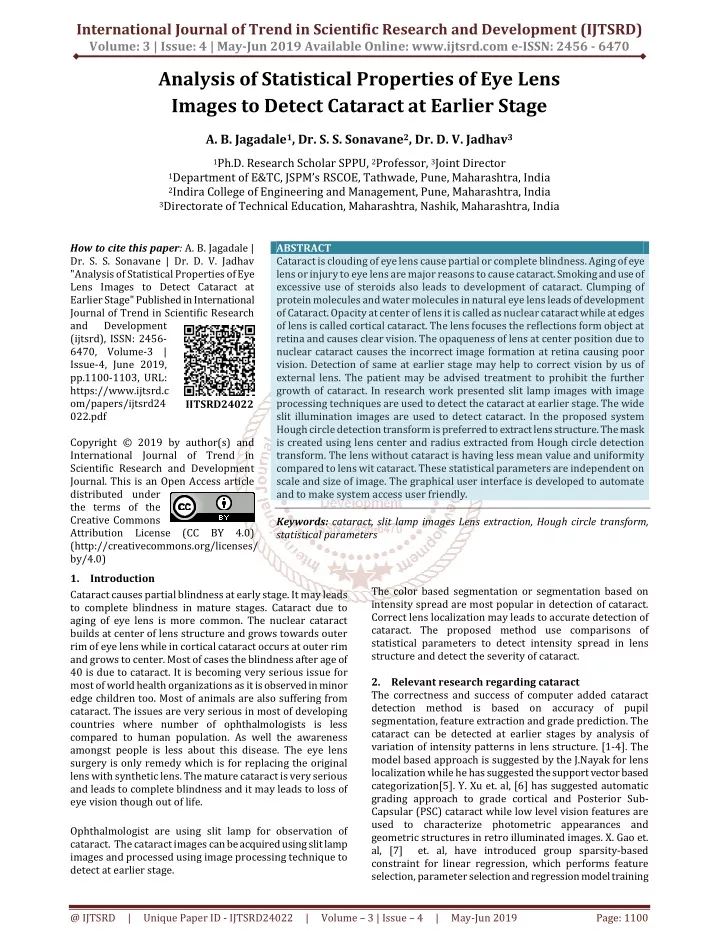
Cataract is clouding of eye lens cause partial or complete blindness. Aging of eye lens or injury to eye lens are major reasons to cause cataract. Smoking and use of excessive use of steroids also leads to development of cataract. Clumping of protein molecules and water molecules in natural eye lens leads of development of Cataract. Opacity at center of lens it is called as nuclear cataract while at edges of lens is called cortical cataract. The lens focuses the reflections form object at retina and causes clear vision. The opaqueness of lens at center position due to nuclear cataract causes the incorrect image formation at retina causing poor vision. Detection of same at earlier stage may help to correct vision by us of external lens. The patient may be advised treatment to prohibit the further growth of cataract. In research work presented slit lamp images with image processing techniques are used to detect the cataract at earlier stage. The wide slit illumination images are used to detect cataract. In the proposed system Hough circle detection transform is preferred to extract lens structure. The mask is created using lens center and radius extracted from Hough circle detection transform. The lens without cataract is having less mean value and uniformity compared to lens wit cataract. These statistical parameters are independent on scale and size of image. The graphical user interface is developed to automate and to make system access user friendly. A. B. Jagadale | Dr. S. S. Sonavane | Dr. D. V. Jadhav "Analysis of Statistical Properties of Eye Lens Images to Detect Cataract at Earlier Stage" Published in International Journal of Trend in Scientific Research and Development (ijtsrd), ISSN: 2456-6470, Volume-3 | Issue-4 , June 2019, URL: https://www.ijtsrd.com/papers/ijtsrd24022.pdf Paper URL: https://www.ijtsrd.com/engineering/computer-engineering/24022/analysis-of-statistical-properties-of-eye-lens-images-to-detect-cataract-at-earlier-stage/a-b-jagadale<br>

E N D
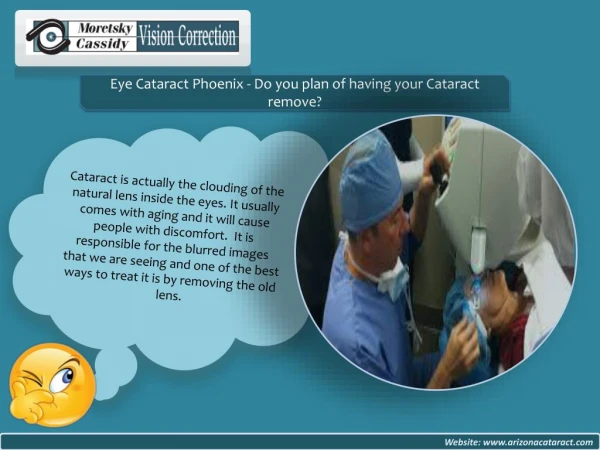
International Journal of Trend in Scientific Research and Development (IJTSRD) Volume: 3 | Issue: 4 | May-Jun 2019 Available Online: www.ijtsrd.com e-ISSN: 2456 - 6470 Analysis of Statistical Properties of Eye Lens Images to Detect Cataract at Earlier Stage A. B. Jagadale1, Dr. S. S. Sonavane2, Dr. D. V. Jadhav3 1Ph.D. Research Scholar SPPU, 2Professor, 3Joint Director 1Department of E&TC, JSPM’s RSCOE, Tathwade, Pune, Maharashtra, India 2Indira College of Engineering and Management, Pune, Maharashtra, India 3Directorate of Technical Education, Maharashtra, Nashik, Maharashtra, India How to cite this paper: A. B. Jagadale | Dr. S. S. Sonavane | Dr. D. V. Jadhav "Analysis of Statistical Properties of Eye Lens Images to Detect Cataract at Earlier Stage" Published in International Journal of Trend in Scientific Research and Development (ijtsrd), ISSN: 2456- 6470, Volume-3 | Issue-4, June 2019, pp.1100-1103, URL: https://www.ijtsrd.c om/papers/ijtsrd24 022.pdf Copyright © 2019 by author(s) and International Journal of Trend in Scientific Research and Development Journal. This is an Open Access article distributed under the terms of the Creative Commons Attribution License (CC BY 4.0) (http://creativecommons.org/licenses/ by/4.0) 1.Introduction Cataract causes partial blindness at early stage. It may leads to complete blindness in mature stages. Cataract due to aging of eye lens is more common. The nuclear cataract builds at center of lens structure and grows towards outer rim of eye lens while in cortical cataract occurs at outer rim and grows to center. Most of cases the blindness after age of 40 is due to cataract. It is becoming very serious issue for most of world health organizations as it is observed in minor edge children too. Most of animals are also suffering from cataract. The issues are very serious in most of developing countries where number of ophthalmologists is less compared to human population. As well the awareness amongst people is less about this disease. The eye lens surgery is only remedy which is for replacing the original lens with synthetic lens. The mature cataract is very serious and leads to complete blindness and it may leads to loss of eye vision though out of life. Ophthalmologist are using slit lamp for observation of cataract. The cataract images can be acquired using slit lamp images and processed using image processing technique to detect at earlier stage. ABSTRACT Cataract is clouding of eye lens cause partial or complete blindness. Aging of eye lens or injury to eye lens are major reasons to cause cataract. Smoking and use of excessive use of steroids also leads to development of cataract. Clumping of protein molecules and water molecules in natural eye lens leads of development of Cataract. Opacity at center of lens it is called as nuclear cataract while at edges of lens is called cortical cataract. The lens focuses the reflections form object at retina and causes clear vision. The opaqueness of lens at center position due to nuclear cataract causes the incorrect image formation at retina causing poor vision. Detection of same at earlier stage may help to correct vision by us of external lens. The patient may be advised treatment to prohibit the further growth of cataract. In research work presented slit lamp images with image processing techniques are used to detect the cataract at earlier stage. The wide slit illumination images are used to detect cataract. In the proposed system Hough circle detection transform is preferred to extract lens structure. The mask is created using lens center and radius extracted from Hough circle detection transform. The lens without cataract is having less mean value and uniformity compared to lens wit cataract. These statistical parameters are independent on scale and size of image. The graphical user interface is developed to automate and to make system access user friendly. Keywords: cataract, slit lamp images Lens extraction, Hough circle transform, statistical parameters IJTSRD24022 The color based segmentation or segmentation based on intensity spread are most popular in detection of cataract. Correct lens localization may leads to accurate detection of cataract. The proposed method use comparisons of statistical parameters to detect intensity spread in lens structure and detect the severity of cataract. 2.Relevant research regarding cataract The correctness and success of computer added cataract detection method is based on accuracy of pupil segmentation, feature extraction and grade prediction. The cataract can be detected at earlier stages by analysis of variation of intensity patterns in lens structure. [1-4]. The model based approach is suggested by the J.Nayak for lens localization while he has suggested the support vector based categorization[5]. Y. Xu et. al, [6] has suggested automatic grading approach to grade cortical and Posterior Sub- Capsular (PSC) cataract while low level vision features are used to characterize photometric appearances and geometric structures in retro illuminated images. X. Gao et. al, [7] et. al, have introduced group sparsity-based constraint for linear regression, which performs feature selection, parameter selection and regression model training @ IJTSRD | Unique Paper ID - IJTSRD24022 | Volume – 3 | Issue – 4 | May-Jun 2019 Page: 1100
International Journal of Trend in Scientific Research and Development (IJTSRD) @ www.ijtsrd.com eISSN: 2456-6470 simultaneously for detection and categorization. R. Supriyanti et.al, [8,9] used specular reflection appearance, texture uniformity and average intensity inside the pupil as cataract detection features. W. Huang et.al, [10] has used neighboring labeled images in a ranked image list, which is achieved using a learned ranking function for grading of nuclear cataract in a slit-lamp image. The ranking function is learned via direct optimization on a newly proposed approximation to a ranking evaluation measure. 3.Methodology The image processing algorithm is developed to detect the cataract using lens localization technique and extracted statistical parameters of lens image. The eye images are acquired using slit lamp mounted camera and processed. From review it is observed that the success of detection of any image processing method depends on the correct and fast localization of lens structure. The lens structure is circular in shape and its color and texture reflects severity of cataract. The lens without cataract is black in color and has mean intensity threshold below 147 for grayscale images with 8 bit resolution. The color based segmentations are popular. But in case of immature and cataract at early stages the detection is hard using color based segmentation. The circular shape of lens encourages the researches to use transform based segmentations. Hough circle detection transform is widely preferred in iris detection and extraction of iris. 4.1 Let lamp in RGB format with 24 bit resolution per pixel. Input image Pre processing is the input color eye image acquired using slit …………(1) Where is gray scale image and , and The input gray scale image is cropped and resized to pixels. It is assumed that the cropped image is containing circular lens and no complete iris. Such that ……………………(2) 4.2 Lens localization using Hough circle detection transform It is assumed that the lens radius is within range of 50 to 65 pixels as image is preprocessed and cropped such that lens radius is within range of 60 to 65. The parameters used in Hough circle detection transform for lower and higher radius range are as below. The Hough transform for circle detection is applied to detect the pupil in image to find out pupil center and radius. The characteristic equation of circle is given by: are green, red and blue planes. ………..(3) Where (a, b) is center of circle and r is radius of circle The circle can be described by two equations: …………………..(4) …………………..(5) Thus the role of hough transform is to search for the triplet of parameters which determines the points . The conversion of image into parametric domain is as show in figure 2 displayed below. Let is the gray scale image obtained from color image . Figure 1 Flow graph for lens localization and feature extraction In proposed method the Hough circle detection transform is used to extract the lens from eye structure. The transform transfers the image from the image space to the parametric space. The accumulator is used to count number of circles passing though given point. The algorithm uses iterative procedure to increment the radius of the circle and store the results. The computational overheads can be reduced by detecting the circle over predefined radius range. Hough circle detection transform returns the center and radius matrix of detected circles. The success of method depends on the detection of only one pupil circle. For same the image is cropped and resized to 120 X 120 pixels to adjust lens radius within range of 60 to 65 pixels range. The extracted pupil center and radius is used to segment the lens structure from eye image. The mean intensity value of the extracted lens without cataract is below 147 and uniformity is below 0.5. These values are much higher for lens with cataract. Figure2. Hough transformation from spatial domain to parametric domain @ IJTSRD | Unique Paper ID - IJTSRD24022 | Volume – 3 | Issue – 4 | May-Jun 2019 Page: 1101
International Journal of Trend in Scientific Research and Development (IJTSRD) @ www.ijtsrd.com eISSN: 2456-6470 4.3 Let image such as mean, homogeneity and smoothness extracted lens are calculated. ∑ j , i Statistical feature extraction: is the extracted lens structure from the eye using Let image For given values of dimensional vector representing the circle parameters where represents the center of circle and r represents radius. Let and are row and column values if input image . is binary image obtained from input gray scale . The statistical parameters and let is the three ..….. (8) = Mean ) j , i ( x ) j , i ( p p(i, j) ∑ j , i = Homogeneit y ...…....(9) + 1 - i j then for all 1 = − Smoothness 1 ∑ j , i …...(10) + 1 ) j , i ( x ……(6) The pupil center and radius is given by such that The mean homogeneity and smoothness are calculated and compared with these parameters of lens without cataract. 5. Result discussions The eye images of 400 volunteers with and without cataract are obtained. The features are calculated and compared. The results are communicated to ophthalmologist and verified from them. The result of ten sample images is displayed as shown in the table below. The proposed systems output and ophthalmologist is compared. It is observed that the results obtained from proposed cataract detection system are correct. Table No. 5.1 Comparison of system results with results of ophthalmologists Image No. Systems Output Doctors opinion Img001 No Img002 Yes Img003 Yes Img004 Yes Img005 Yes Img006 No Img007 No Img008 Yes Img009 Yes Img010 Yes Table 2 indicates the confusion matrix generated to calculate sensitivity, specificity and accuracy obtained. ………….(7) The lens extracted using Hough circle detection algorithm is as displayed in figure 3. Healthy / N early / Y Nuclear / Y Nuclear /Y mature / Y Healthy / N Cortical / Y Nuclear/ Y early/ Y early / Y Figure3. Lens extracted from slit lamp eye image without cataractThe image displays the original image, lens detected, mask for detected lens and extracted lens. Table 2: Confusion matrix and results for Hough Circle Detection transform and correlation Predicted Cataract by proposed algorithm Yes No Yes 200 60 No 40 100 Actual observation Total Number of Images Cataract Affected Sensitivity Specificity Accuracy 400 240 75 62.25 83.33 The accuracy indicated by above method is 83.33% for set of 400 images. The GUI for detection of cataract has been developed using MATLAB. The images obtained from government hospital Pandharpur have been processed using Hough circle detection transform and correlation. The result of cataract detection of eye without cataract is as displayed in figure 3. @ IJTSRD | Unique Paper ID - IJTSRD24022 | Volume – 3 | Issue – 4 | May-Jun 2019 Page: 1102
International Journal of Trend in Scientific Research and Development (IJTSRD) @ www.ijtsrd.com eISSN: 2456-6470 [3]The lens opacities classification system III, L. T. Chylack, J. K. Wolfe, D. M. Singer, M. C. Leske, et al, , Archives of Ophthalmology, 111 (1993) 831-836. [4]Assessment of Cataracts from Photographs in the Beaver Dam Eye Study, B. E. K. Klein, R. Klein, K. L. P. Linton, Y. L. Magli, M. W. Neider, Ophthalmology, 97, 11 (1990) 1428-1433. [5]Automated Classification of Normal, Cataract and Post Cataract Optical Eye Images using SVM Classifier, J.Nayak Proceedings of the World Congress on Engineering and Computer Science 2013, 1 (2013). [6]Automatic Grading of Nuclear Cataract from Slit-Lamp Grading Images Using Group Sparsity Regression Y. Xu, X.Gao, S.Lin, D.Wing, K.Wong, J.Liu, D.Xu, C.Y. Cheng, C.Y. Cheung, T.Y.Wong , Medical Image Computing and Computer-Assisted Intervention-MICCAI 2013, Lecture Notes in Computer Science, 8150 (2013) 468-475. Figure 3 Detection for eye without cataract 6. Conclusion Cataract causes lens opacity which leads to partial or complete blindness. The automated image processing techniques can be used to develop system to detect the cataract at earlier stage and help ophthalmologist to minimize inter grader and intra-grader errors. The Hough circle detection transform used is accurate to detect the lens structure gives lens radius and centers used for lens extraction. The statistical homogeneity and smoothness are used to differentiate lens with cataract and without cataract. The cataract detection accuracy is 83.33%. 7. References [1]Prevalence of cataract and pseudophakia/aphakia among adults in the United States, The Eye Diseases Prevalence Research Group, Archives of Ophthalmology, 122 (2004) 488-494. [7]Automatic Grading of Cortical and PSC Cataracts Using Retroillumination Lens Images, X. Gao, D.W.K. Wong, T. T. Ng, C.Y. L. Cheung, C.Y. Cheng, T. Y. Wong, Computer Vision –ACCV 2012, Lecture Notes in Computer Science, 7725 (2013) 256-267. [8]Consideration of Iris Characteristic for Improving Cataract Screening Techniques Based on Digital Image R. Supriyanti, Y. Ramadhani, 2012 2nd International Conference on Biomedical Engineering and Technology IPCBEE, 34 (2012) 126-129. parameters like mean, [9]The Achievement of Various Shapes of Specular Reflections for Cataract Screening System Based on Digital Images, R. Supriyanti, Y. Ramadhani, 2011 International Conference on Biomedical Engineering and Technology IPCBEE, 11 (2011) 75-79. [10]A Computer Assisted Method for Nuclear Cataract Grading From Slit-Lamp Images Using Ranking, W. Huang, K. L .Chan, H. Li, J. H. Lim, J. Liu, And T. Y. Wong, IEEE Transactions On Medical Imaging, 30, 1(2011) 94- 107. [2]The World Health Report: Life in the 21st Century – A Vision for All, World Health Organization, Geneva, 1998. @ IJTSRD | Unique Paper ID - IJTSRD24022 | Volume – 3 | Issue – 4 | May-Jun 2019 Page: 1103