Download
1 / 15
150 likes | 277 Views
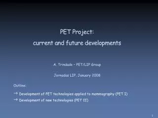
PET Project: current and future developments. A. Trindade – PET/LIP Group Jornadas LIP, January 2008. Outline: Development of PET technologies applied to mammography (PET I) Development of new technologies (PET II). Breast Cancer and PET.

E N D
PET Project:current and future developments A. Trindade – PET/LIP Group Jornadas LIP, January 2008 Outline: • Development of PET technologies applied to mammography (PET I) • Development of new technologies (PET II)
Breast Cancer and PET • Breast cancer is the most common form of cancer among women: 4500 new cases each year in Portugal • Whole body PET using 18F-FDG radiotracer has an increasing application for breast cancer detection PET: physiological functions of the tissue X-ray: lesion size, shape and tissue density infiltrating carcinoma L. P Adler, Fox Chase Cancer Center There is a need to improve the sensitivity and specificity of PET for small lesions
Breast exam Axilla exam Positron Emission Mammography (PEM) • Dedicated PET scanners for breast imaging • ClearPEM requirements: • High detection sensitivity • Spatial resolution (1-2 mm FWHM) • Time resolution for backgr. rejection (1-2 ns) • Shorter exams and/or less dose (370 MBq) • Detector concept: • Two planar heads (FOV ~ 15 cm x 17 cm) • Mammary gland and axilla region exams • Exam with the patient in prone position • Ajustable distance between heads and rotation angle
The PET-Mammography Consortium The PET-Mammography consortium was created in December 2002 LIP (Lab. Instrumentação e Física Exp. de Partículas) IBEB (Inst. Biofísica e Engª Biomédica - FCUL) IBILI (Inst. Biomédico Invest. Luz e Imagem - FMUC) 8 Institutes 40 People Crystal Clear Collab. INOV / INESC-ID (Inst. Engª Sistemas Computadores) INEGI (Inst. Engª Mecânica e Gestão Industrial) HGO (Hospital Garcia de Orta) TagusPark S.A. Financiamento: AdI (Agência de Inovação)
PET/LIP Group R. Bugalho, B. Carriço, M. Ferreira, R. Moura, C. Ortigão, J. Pinheiro, J. C. Silva, P. Rodrigues, A. Trindade, J. Varela • Scientific coordination • R&D activities on radiation detectors • Electronic systems arquitecture and development • Data acquisition software development • Detector design and performance studies • Detector integration • Commissioning and pre-clinical tests
Single readout (flood irradiation) Detector Module Performance LIP • Optimization studies: • Crystal surface roughness • Reflector type • Optical coupling medium • Characterization studies: • Energy resolution: 15% @ 511 keV • Ligth collection • DOI resolution: 2.2 mm FWHM • Cross-talk contamination: 4-5% • Ageing effects • Discrete 32 channels electronic setups Double readout (flood and colimated irradiations)
APD Quality Control LIP • 400 Hamamatsu APD quality control • Gain vs bias voltage • Dark current • Gain gradient • One person full time for 4 months (2006) • Development of an automated setup (2007): • Slots for 16 APDs • Settings - Altera DE2 board/Labview • 10 days for 400 APDs
Cooling System (working Temp.=18ºC) Service Rack Control Rack Detector Heads Scanner Operator Workstation 1. Robot Data Acquisition Electronics (L1 hardware trigger) 2. Acquisition tool (LIP, IBEB, IBILI) 3. INEGI 4. Detector and data info. 5. INEGI Reconstruction and visualization software 1. FE Emulator Electronics (LIP, INOV) 2. DAE Server (L2 software trigger) (LIP) 3. Service Manager (slow control sw) (LIP) 4. High Voltage Supplys 5. Low Voltage Supplys ClearPEM at Hospital Garcia de Orta (Installation in 2008, clinical trials with 300 patients)
First ClearPEM Image (Na-22 source) LIP, IBEB Acquired Data Simulated Data
Lesion Detectability (5 min. exam) LIP, IBEB • Results support that ClearPEM scanner can improve • the detection of breast lesions • 5 mm lesions: 100% detectability for all L/B FDG ratios • 3 mm lesions: 100% detectability for the highest L/B FDG ratios 3D-OSEM images of a breast phantom from simulated data Images not corrected for attenuation, scatters or randoms
PET I Activities • Detector Integration • First full integration exercice of the ClearPEM system: May 2007 at TagusLIP • All the sub-systems were tested (next talk): • Detector heads integration • Front-End electronics • Data acquisition electronics • Data acquisition and slow control software • Some revisions are currently in progress before the final integration • Commissioning and Pre-clinical tests • Detector calibration methods • Image correction techniques: normalization • Tests with radioactive phantoms • Definition of the scanner operation protocols
First floor Laboratory Area First floor Bunker for work with radioactive sources Inside the bunker Computing TagusLIP: LIP Laboratory at TagusPark • Reorganization of the laboratory in 2007 • Internal network upgraded to Gigabit • Dedicated servers for data analysis and electronics developments • Mass storage in NFS server • Automatic synchronization with PET storage dCache area (FTP Grid)
PET II Activities • New PET detector modules • Revised APD array / evaluation of Silicon Photomultipliers (SiPMTs) • Revised electronics: frontend and off-detector data acquisition (next talk) • Small animal imaging platform • ClearPEM-Sonic (PET-US) • PET insert for Magnet Resonance Imaging (PET-MRI)
Baseline design like ClearPEM but several upgrades foreseen To be installed at TagusLIP: A technological demonstrator for future PET R&D developments Link with institutes interested in PET for biomedical applications Moby (mouse) phantom Planar geometry Small Animal Imaging Platform
Small Animal Images Phantom Geometry Anatomy/Activity Fusion Soft tissue in gray Bone squeleton in white Bone uptake Reconstructed Activity LIP, IBEB 3D-OSEM images of a mouse bone scan (18F) from simulated data