Download

1 / 18
240 likes | 1.02k Views
Treatment of Periocular Necrotizing Fasciitis and Mucormycosis with Surgical Debridement and Hyperbaric Oxygen Therapy. Yang, Paul Chung, Ji Won Song, Julia MD Song, Alice MD April 9-14, 2010 ASCRS/ASOA Boston, MA Authors have no financial interest. Introduction Necrotizing fasciitis .

E N D
Treatment of Periocular Necrotizing Fasciitis and Mucormycosis with Surgical Debridement and Hyperbaric Oxygen Therapy Yang, Paul Chung, Ji Won Song, Julia MD Song, Alice MD April 9-14, 2010 ASCRS/ASOA Boston, MA Authors have no financial interest
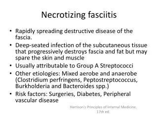
IntroductionNecrotizing fasciitis • Rare opportunistic subcutaneous infection. • Affects subcutaneous soft tissue of trunk, extremities and perineum. • Infection in orbit threatens individual’s vision, cavernous thrombosis leads to death. • Infection leads to fatal consequences if not treated earlier.
IntroductionRhinocerebral mucormycosis • Opportunistic infection caused by saprophytic fungi. • Most common in diabetic and immunocompromised individuals. • Infection starts at paranasal sinus and rapidly spreads to orbit, cavernous sinus and brain • Causes thrombosis, localized acidosis, tissue ischemia and necrosis. • Spread in CNS could cause death.
PathophysiologyNecrotizing fasciitis • Caused by group A and non-group A streptococcus and staphylococcus. • Infection rapidly spreads through tissues; vascular thrombosis and tissue necrosis follow. • Orbital involvement of infection causes eyelid ischemia, necrosis, ophthalmic artery and even death. • Shows high resemblance to cellulitis, sinusitis and mucormycosis.
PathophysiologyRhinocerebral mucromycosis • Fungal infection caused by microorganisms in genera Mucor, Absidia and Rhizopus. • Normally present on respiratory tract mucosa and paranasal sinuses as airborne spores, turn pathogenic if individual suffers from immunological and metabolic disorders. • Infected individual may complain of facial pain, proptosis, and ophthalmoplegia. From thrombosis, acidosis, tissue ischemia. • Infection into CNS could cause death.
Method • This presentation introduces successful treatment of each bacterial/fungal infection involving surgical debridement and adjunct hyperbaric oxygen therapy. • Case 1: Necrotizing fasciitis • Case 2: Rhinocerebral mucormycosis
Pain with Eye Edema (+/- Sinusitis) Evaluation & Management • Exam • MRI, Nuclear medicine • Lab (Glucoses, cultures, etc) • History • CBC • X-ray Necrotizing fasciitis Rhinocerebral mucormycosis • Surgical Debridement of sinuses and orbit • Antifungal Agents • Hyperbaric Oxygen Treatment • Surgical Debridement • IV Antibiotics • Hyperbaric Oxygen Treatment
CASE 1: Necrotizing fasciitis • 41 year-old patient came to the Long Beach Memorial Medical Center emergency room with pain, swelling and discharge from left medial canthal region. • VA: 20/20 OD and 20/40 OS. Intraocular pressures: 13 OD and 22 OS mm Hg. • Extraocular motility was full in OD while in OS, it decreased to -1. • Ptosis and 4 + facial edema were present on the left orbit. Preoperative : Extensive upper and lower eyelid edema. MRI: Marked perioribital involvement (edema)
Case 1: Necrotizing fasciitis • IV antibiotocs (Vancomycin, Piperacillin/tazobactam and Levofloxacin). • Surgical debridement of the infected tissues. • Drain was left in place for 2 days and hyperbaric oxygen chamber was started next day for 2 atm for 90 minutes for 9 treatment sessions. • Patient was discharged on the 5th day on oral antibiotics. Intraoperative: Ischemia and edema of orbicularis muscle with removal of exudative material with a curette. Immediately postoperative with drain in place. One week post-op with reduction of erythema and edema
Case 1: Necrotizing fasciitis Several months later without need for reconstruction.
CASE 2: Rhinocerebral mucormycosis Patient with rhino-orbital mucormycosisprior to surgery. • Retrospective chart review of rhino-orbito or cerebral mucormycosis patients at Loma Linda University Medical Center, CA from 2000 to 2005. Orbital CT scan with mucormycosisinvolving ethmoid sinus and orbit.
CASE 2: Rhinocerebral mucormycosis • Infected patients had following predisposing factors: uncontrolled, diabetes, immunosupression after chemotherapy and due to prednisone intake. • Most patients complained of Facial pain and displayed periorbital edema. • Maxillary sinus was most frequent infection area, followed by ethmoid and sphenoid. Frequency of Signs and Symptoms Frequency of Sinus Involvement
CASE 2: Rhinocerebral mucormycosis • Surgical debridement of infected tissue, extensive sinus surgery followed by oribtal exenteration if necessary. • Amphotericin B and Hyperbaric oxygen treatment. Patient with rhino-orbital mucormycosis s/p anterior orbitotomy with a Penrose drain.
CASE 2: Rhinocerebral mucormycosis Patient did well postoperatively without visual problems.
Results • Case 1: After administering intravenous antibiotics and surgical debridement of infected tissues followed by adjunct Hyperbaric Oxygen treatment for 2 weeks, the individual improved significantly without vision loss and no need for exenteration or reconstruction.
Results • Case 2: Successful treatment of mucormycosis involves surgical debridement of infected tissues followed by Amphotericin B. Also, managing predisposing factors for infection played important role in treating mucormycosis. Adjunct hyperbaric oxygen treatment improved patients recovery as well.
Mechanisms for Hyperbaric Oxygen Treatment • 1. Oxygenation of Ischemic, Hypoxic Tissue - HBO increases plasma and tissue oxygen tensions 10 times which helps tissues to maintain tissue viability without RBC’s • 2. Neutrophil Oxidative Killing - Neutrophils regain ability to kill bacteria by generation of oxygen dependent superoxides and peroxides. • 3. Suppression of multiplication in bacteria- High oxygen tensions suppress growth of streptococcus • 4. Augmentation of Antibiotic effectiveness- Increased oxygen tension allows oxygen dependent active transport to bring in antibiotic across bacterial cell wall. • 5. Enhanced fibroblast function- prevents unnecessary cicatrix formation. • 6. Angiogenesis- Increased oxygen tension increases vascular endothelial grow factor function as well as secretion of matrix by fibroblasts
Conclusion • Periorbital necrotizing fasciitis can be managed with prompt, aggressive treatment and adjunctive hyperbaric oxygen therapy; its complications can be controlled as well. Our case 1 patient recovered completely with satisfactory cosmesis and minimal cicatricial changes. • Rhinocerebral mucormycosis is an infection that could be treated with aggressive surgical treatment, mainly debridement, exenteration, and followed by systemic fungal therapy. Identification of predisposing factors and managing them can aid in rapid diagnosis of patients at high risk and rate of survival. • Adjunctive hyperbaric oxygen treatment complements surgical interventions and antibiotics in this potentially vision losing, loss of life conditions and help infected individuals recover.