Download

1 / 19
210 likes | 625 Views
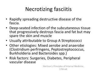
Necrotizing fasciitis. Necrotizing fasciitis. Diagnosis?- Necrotizing fasciitis What is the most common predisposing factor? A. Trauma B. Surgery C. Vascular abnormality D . . What is the hallmark finding? A. Edema B. Swelling C. Soft tissue gas D. Cortical Erosion.

E N D
Necrotizing fasciitis • Diagnosis?- Necrotizing fasciitis • What is the most common predisposing factor? • A. Trauma • B. Surgery • C. Vascular abnormality • D.
What is the hallmark finding? • A. Edema • B. Swelling • C. Soft tissue gas • D. Cortical Erosion
DIABETIC MUSCLE INFARCTION • 45 year old malewith a history of uncontrolled diabetes. One day history of left thigh pain aggravated by movement.
DIABETIC MUSCLE INFARCTION • Diagnosis? • Most commonly affected muscle? • A. Thigh • B. Forearm • C. Arm • D. Hand • Imaging of choice? • A. X-ray - C. MRI • B. Ultrasound - D. CT scan
MyositisOssificans • Diagnosis? • Most important radiographic manifestation? • A. rim calcification and ossification with a lucent center • B. Cortical erosion • C. Soft tissue swelling • D. Sunburst reaction
This usually is seen after how long? • A. 2-3 weeks • B. 2-3 months • C. 5 to 6 months • D. 2-3 days
Compartment syndrome • Diagnosis? • Contrast enhancement is used for?- • A.distinguishingbetween perfused and devitalized muscle. • B. determine other possible pathology. • C. distinguished thrombus formation • Most common site? • A. volar compartment of the forearm • B.anteriorand deep posterior compartment of the leg • C. All of the above • D. None of the above.
Benign Gastric ulcer • Diagnosis • Hallmark of benign ulcer? • A.Mucosathat is intact to the very edge of an undermining ulcer crater • B. Eccentric location within the lumen of the stomach • C. Width greater than depth • D. Nodular , rolled, irregular, or shouldered edges • A large flat-based ulcer with heaped-up edges that fold inward to trap a lens shaped barium collection that is convex toward the lumen?
Pseudomembranous colitis • Most probable diagnosis? • Contributing causes except? • A. surgery • B. Antibiotics • C. intestinal ischemia • D. irrdiation • Characteristic CT finding? • A. • Accordion sign
duodenitis • Diagnosis • Major Cause? • A. H. Pylori infection • B. Alcohol • C. anti-inflammatory medications • UGI findings except • A. Thickening of proximal duodenal folds • B. Enlarged Brunner glands • C. Surrounding fat strandings • D. Deformity of the duodenal bulb
Infectious esophagitis • Diagnosis? • Most common cause of this disease? • A.Candidaalbicans • B. Herpes simplex • C. tuberculosis • D. Cytomegalovirus • Radiographic findings include • A. Stricture formation • B. Abscess formation • C.Tinynodular, or they may be giant and coalescent with pseudomembranes • D. Sinus tract formation