Download
1 / 41
430 likes | 533 Views
Immunolocalization for EM. Using immunoglobulin molecules as tags for select proteins and carbohydrates. Visualized by using colloidal gold or enzyme reactions. Leishmania megasome labeled with 10nm gold. Immune Responses. 1. Humoral :

E N D
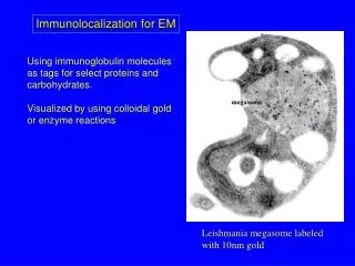
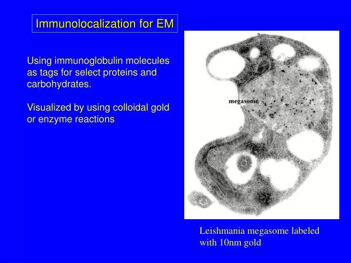
Immunolocalization for EM Using immunoglobulin molecules as tags for select proteins and carbohydrates. Visualized by using colloidal gold or enzyme reactions Leishmania megasome labeled with 10nm gold
Immune Responses 1. Humoral: B lymphocytes produce antibodies recognizing an antigen from foreign substance. Antibodies are then secreted into blood stream.
Immune Responses 1. Humoral: B lymphocytes produce antibodies recognizing an antigen from foreign substance. Antibodies are then secreted into blood stream. 2. Cell-mediated: Mature T lymphocytes - antigen responding, response control, and response mediating cells
Immunoglobins (Ig) IgG IgA
Immunoglobins (Ig) IgG IgA IgM
Glossary Antibody (anti-foreign body) is a protein produced by a white cell (B lymphocyte).
Glossary Antibody (anti-foreign body) is a protein produced by a white cell (B lymphocyte). Antigen (antibody generating substance) is any agent, such as a chemical or microorganism that is recognized by the antibody. Not all antigens are immunogens (e.g hapten).
Glossary Antibody (anti-foreign body) is a protein produced by a white cell (B lymphocyte). Antigen (antibody generating substance) is any agent, such as a chemical or microorganism that is recognized by the antibody. Not all antigens are immunogens (e.g hapten). Immunogen : Any substance to which an animal responds by making antibodies. All immunogens are antigens.
Glossary Antibody (anti-foreign body) is a protein produced by a white cell (B lymphocyte). Antigen (antibody generating substance) is any agent, such as a chemical or microorganism that is recognized by the antibody. Not all antigens are immunogens (e.g hapten). Immunogen : Any substance to which an animal responds by making antibodies. All immunogens are antigens. Antigen binding site - relatively small region of an antibody that binds to the antigen.
Epitope (antigenic determinant) - is that part of an antigen that is recognized by a single antibody.
Epitope (antigenic determinant) - is that part of an antigen that is recognized by a single antibody. Hapten - low molecular weight compounds (such as plant hormones) that typically do not elicit a spontaneous immune response but can be recognized by antibodies. Typically attached to an immunogen.
Epitope (antigenic determinant) - is that part of an antigen that is recognized by a single antibody. Hapten - low molecular weight compounds (such as plant hormones) that typically do not elicit a spontaneous immune response but can be recognized by antibodies. Typically attached to an immunogen. Hybridoma - fusion product between B cell and myeloma cell (“immortal cell”).
Epitope (antigenic determinant) - is that part of an antigen that is recognized by a single antibody. Hapten - low molecular weight compounds (such as plant hormones) that typically do not elicit a spontaneous immune response but can be recognized by antibodies. Typically attached to an immunogen. Hybridoma - fusion product between B cell and myeloma cell (“immortal cell”). HAT selection - culture media that contains hypoxanthine, aminopterin and thymadine. A selective media that only allows hybridomas to grow.
Terms used in Immunolabeling Primary antibody: An antibody that is specific to the antigen of the sample to be localized. Can be conjugated to a signal (gold, fluorochrome or enzyme).
Terms used in Immunolabeling Primary antibody: An antibody that is specific to the antigen of the sample to be localized. Can be conjugated to a signal (gold, fluorochrome or enzyme). Secondary antibody: An antibody that recognizes a primary antibody. Usually always is conjugated to signal.
Terms used in Immunolabeling Primary antibody: An antibody that is specific to the antigen of the sample to be localized. Can be conjugated to a signal (gold, fluorochrome or enzyme). Secondary antibody: An antibody that recognizes a primary antibody. Usually always is conjugated to signal. Diluent: Physiologic buffer and non-specific protein (e.g. albumin or non-fat milk) used in diluting the antibodies. Sometimes detergent added to decrease surface tension of sections.
Block: Physiologic buffer, high salt, and non-specific protein. The protein adheres to any “sticky” sites that might allow non-specific binding of antibodies.
Block: Physiologic buffer, high salt, and non-specific protein. The protein adheres to any “sticky” sites that might allow non-specific binding of antibodies. Etching: treating resin sections with HCl or sodium borohydride to reduce steric hindrance or expose hidden antigenic sites.
Antibody Production Polyclonal: Antibodies are collected from sera of exposed animal, - or - a combination of monoclonal colonies is combined.
Antibody Production Polyclonal: Antibodies are collected from sera of exposed animal, - or - a combination of monoclonal colonies is combined. Can be any animal: Rabbit, Goat, Horse, Rat, Sheep, etc…
Antibody Production Polyclonal: Antibodies are collected from sera of exposed animal, - or - a combination of monoclonal colonies is combined. Can be any animal: Rabbit, Goat, Horse, Rat, Sheep, etc… Suite of antibodies recognizing multiple antigenic sites of injected biochemical.
Monoclonal: Individual B lymphocyte hybridoma is cloned and cultured. Secreted antibodies are collected from culture media.
Monoclonal: Individual B lymphocyte hybridoma is cloned and cultured. Secreted antibodies are collected from culture media. Typically BALBc mice Sometimes Rat (ascites fluid).
Monoclonal: Individual B lymphocyte hybridoma is cloned and cultured. Secreted antibodies are collected from culture media. Typically BALBc mice Sometimes Rat (ascitesfluid). Antibodies recognize one antigenic binding site of the antigen.
Determining which B cell to clone ELISA or Immuno-fluorescence
Generic TEM Immunolabeling Protocol Fixation: Glutaraldehyde only Dehydration Embedding in methacrylate resin (e.g. LR White, Lowicryl, or Quetol) Section Immunolabel Optional - Post-stain
Controls Adsorption - primary antibody exposed to excess of antigen to remove any labeling. Label without antibody - no antibody, shows labeling not due to reaction with label Omission of primary antibody - the secondary or tertiary antibodies should not recognize the tissue. Pre-immune sera - collected from animals that have not produced antibodies, or use “normal serum” from non-immunized animals of same species
Immunolabeling Sections - Float grids on blocking solution - Incubate in primary antibody - Wash thoroughly with buffer to remove unbound antibody
Incubate with secondary antibody or protein A/G Wash again to remove unbound secondary Last wash should be water.
Double Labeling Incubate sections with second primary antibody of interest
Incubate with secondary - different size gold Note steric hinderence at asterisk
How are the antibodies coupled to the gold?The proteins are attached to the gold particles by: • a). Charge attraction (Lysine) • b). Hydrophobic attraction (Tryptophan) and • c). Sulfur binding (Cysteine and Methionine) Strongest binding suggested to be the sulfur "hinge" joining the the two Fc regions
Using Protein A or Protein G instead of secondary antibody
Ferritin enhanced labeling Silver enhanced
Freeze fracture immunolabeled
Freeze fracture immunolabeled Negative stain Immunolabeled