Download
1 / 38
380 likes | 557 Views
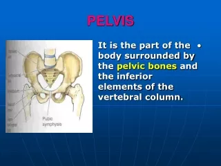
Pelvis and Contents. Reproductive Organs and System. Bony Pelvis. 2 Pelvic = Coxal = Innominate bones fused together Each Pelvic bone Ilium Ischium Pubis 3 parts join to form acetabulum Sacrum and Coccyx help create pelvis and form pelvic cavity Function

E N D
Pelvis and Contents Reproductive Organs and System
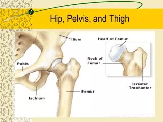
Bony Pelvis • 2 Pelvic = Coxal = Innominate bones fused together • Each Pelvic bone • Ilium • Ischium • Pubis • 3 parts join to form acetabulum • Sacrum and Coccyx help create pelvis and form pelvic cavity • Function • attaches lower limb to axial skeleton • supports viscera • transmits weight of upper body Use lab work to learn bony landmarks of pelvis Pg 187
Contents of Pelvic Cavity • True Pelvis • below pelvic brim • space contains • part colon • rectum • bladder • uterus/ovaries (females) • False Pelvis • iliac blades • above pelvic brim • contains abdominal organs • attachment for muscles + ligaments to body wall • Pelvic Diaphragm = levator ani + coccygeus m
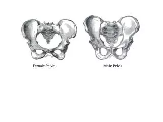
Cavity is broad, shallow Pelvic inlet oval + outlet round Bones are lighter, thinner Pubic angle larger Coccyx more flexible, straighter Ischial tuberosities shorter, more everted Cavity is narrow, deep Smaller inlet + outlet Bones heavier, thicker Pubic angle more acute Coccyx less flexible, more curved Ischial tuberosities longer, face more medially Sexual Dimorphism in Pelvis Female Male
Sexual Dimorphism in Pelvis pg 189
Perineum • Diamond-shaped area between • Pubic symphysis (anteriorly) • Coccyx (posteriorly) • Ischial tuberosities (laterally) • Males contain • Scrotum, root of penis, anus • Females contain • External genitalia, anus pg 744
Development of Reproductive Organs • Gonadal ridge: Forms in embryo at 5 weeks Gives rise to gonads Male gonads = testis Female gonads = ovaries
Reproductive Embryology • Male and Female ducts are both present in early embryo, but only one set develops! • Wolffian ducts (Mesonephric): form male ducts • vas deferens, epididymis • Mullerian ducts (Paramesonephric): form female ducts • uterus, oviduct, vagina
External genitalia develops from same structures • Embryonic structure Male Female • Labioscrotal swelling Scrotum Labia major • Urethral folds Penile Urethra Labia minor • Genital tubercle Penis Clitoris
Male Development • Male fetus • Testes descend partially at 3 months, finish at 7 months into scrotum • Vaginal Process: outpocketing of peritoneum forms tunica vaginalis • Gubernaculum: fibrous cord; attaches bottom of scrotum to testes • Testes Descent: partly due to shortening of gubernaculum, final descent due to testosterone and maybe increase in intra-abdominal pressure
Female Development • Ovaries descend into pelvis • Vaginal process: outpocketing of peritoneum guides descent • Gubernaculum:guides descent of ovaries; attached to labia major • caudal portion = round ligament of uterus • cranial portion = ovarian ligament
Females = around 11 breasts enlarge increase subcutaneous fat in hips and breasts hair in pubic and axillary region oily skin menstruation (1-2 years later) Males = around 13 scrotum + testes enlarge enlargement of larynx increase in body size, musculature hair in facial, pubic, axillary regions oily skin Puberty: period where reproductive organs grow and can reproduce
Reproductive SystemGenitalia = sex organs Primary = ovaries, testesSecondary = glands, ducts, external genitalia Female Male pg 5
Male Reproductive System • Primary Sex Organs • testes • Accessory Sex Organs • External Genitalia • penis • scrotum • Ducts • Efferent ductules (epididymis) • vas deferens • ejaculatory duct • urethra • Glands • seminal vesicle • prostate • bulbourethral pg 672
Male Reproductive Anatomy • Scrotum • sac of skin + superficial fascia • contains testes • Associated Muscles • Dartos: inside skin of scrotum • wrinkles skin = warm • Cremaster: extends into scrotum from spermatic cord • Fibers from internal oblique • elevates testes = warm • lower testes = cool • Tunica vaginalis = light sac • covering each testis • Tunica albuginea = fibrous • deep to tunica vaginalis • divides testes into lobules pg 673
Male Reproductive Anatomy: Testes • Seminiferous Tubules • make-up testes • location of spermatogenesis • Divided into lobules • Tubulus Rectus • convergence of seminiferous tubules • Rete Testis • network of branching tubes • leads to epididymis pg 674
Male Reproductive Anatomy: pg 704 • Epididymis • Contains efferent ductules: tube from rete testis to duct of epididymis • gain ability to swim here • smooth muscle layer = ejaculation • epithelial layer lined w/stereocilia • resorb excess testicular fluid • transfer nutrients to sperm in lumen • Vas Deferens • tube from duct of epididymis to ejaculatory duct • Vasectomy-cut vas deferens, close off end pg 672, 674
Cell Division • Mitosis: cell division with chromosome duplication and division 2 daughter cells = parent • Have Diploid = 2n number of chromosomes • Occurs in body (somatic) cells • Meiosis = Reduction Division: cell division resulting in cells having half the number of chromosomes as parent • Have Haploid = n number of chromosomes • Occurs in sex cells
Spermatogenesis: production of sperm • Stem cells = Spermatogonia (2n) • Undergo Mitosis • Type A spermatogonia = precursor cells (2n) • Type B spermatogonia = primary spermatocytes (2n) • Primary spermatocytes undergo Meiosis I 2 secondary spermatocytes (n) • 2 Secondary spermatocytes (n) undergo Meiosis II 4 spermatids (n) • Spermiogenesis: maturation of spermatids into spermatozoa (sperm) • Head (acrosome), midpiece, tail • Controlled by FSH (pituitary gl.), Testosterone (testes)
Within Seminiferous Tubules • Sustenacular (Sertoli) cells: surround spermatogonia in lumen of seminiferous tubules • Provide nutrients to spermatogenic cells • Move cells toward tubule lumen • Secrete testicular fluid • Phagocytize cytoplasm shed by developing spermatids • Secrete Androgen-binding protein (concentrates testosterone) • Secrete Inhibin: hormone slows rate of sperm production • Blood-testis barrier: sustenacular cells bound together by tight junctions to prevent escape of membrane antigens from sperm into blood • Myoid Cells: layer around seminiferous tubules of smooth muscle • Interstitial (Leydig) Cells: in loose CT between seminiferous tubules secrete androgens (male sex hormones)
Spermatic Cord Collective name for structures associated with the scrotum • Passes through inguinal canal • Includes • Vas Deferens • Testicular Arteries + Veins • Lymphatic vessels • Cremaster muscle fibers • Nerves pg 673
AccessoryGlands • Seminal vesicle (paired) • posterior surface of bladder • contracts during ejaculation • empties into vas deferens • Functions • nourish sperm • stimulate uterine contractions • suppress immune response • enhance sperm motility • clot ejaculated semen once in vagina, then liquefy sperm to allow swim • Prostate • inferior to bladder, anterior to rectum • encircles first part of urethra • contracts during ejaculation • Functions: clot, liquefy, motility pg 672
Accessory Glands pg 672 • Bulbourethral (paired) • inferior to prostate • within urogenital diaphragm • empties into spongy urethra • Function: produce mucous • neutralize urine in urethra • lubricate semen for passage
Penis • Male external genitalia • Function: delivers sperm into the female reproductive tract • Anatomy • root = attached end • crura-anchored to pubic arch, covered by ischiocavernosus muscle • bulb-secured to urogenital diaphragm • shaft/body = free, not attached • glans penis = enlarged tip • prepuce = loose cuff around glans • spongy urethra = tube within penis pg 680
Penis (continued) • Erectile bodies • 3 long strips of erectile tissue around the spongy urethra • thick tube covered by dense CT and filled with smooth muscle, CT + vascular spaces • Corpus spongiosum • distally = glans penis • proximally =bulb of penis • midventral erectile body • Corpora cavernosa • proximally = root/crura of penis, covered by ischiocavernosus m. • paired, dorsal erectile bodies • make up most of mass pg 680
Penis (continued) • Arterial Supply = branches of Internal Pudendal (branch of internal iliac) • Innervation = branches of Pudendal (from sacral plexus) provide sensory • Parasympathetic: engorgement of blood in erectile bodies = erection • Sympathetic: contraction of smooth muscle in ducts and glands and bulbospongiosum m = ejaculation • Above Autonomic from inferior hypogastric plexus
Female Reproductive System • Primary Sex Organs • Ovaries = gonads • Accessory Sex Organs • External Genitalia = vulva • Labia major + minor • Mons pubis • Clitoris • Ducts • Uterine tube = oviducts • Vagina • Glands • Greater vestibular gland pg 684
Female Reproductive Anatomy • Ovaries (paired) • produce and store ova (eggs) • Produce estrogen • Tunica albuginea - surrounds each ovary • Germinal epithelium-external to tunica albuginea (= mesothelium) • Arterial Supply • Ovarian & branches of uterine a. • Ligaments • Ovarian ligament • connects ovaries to uterine wall (medial) • Suspensory ligament • connects ovaries to pelvic wall (lateral) • Broad ligament • supports uterus, oviducts • Round Ligament (part of broad) • Attaches uterus to labia majorum pg 685
Oogenesis: production of eggs (ova) • Stem cells = oogonia undergo Mitosis • all of female’s oogonia produced while fetus • Oogonia begin Meiosis I are called primary oocytes (2n) • Meiosis I is stalled before birth • During ovulation, Meiosis I completed and Meiosis II begins • Once Meiosis II begins, primary oocytes now called secondary oocytes (n) • Meiosis II is completed when sperm penetrates egg • When Meiosis II is completed, secondary oocyte is now called ovum (egg) • Meiosis II results in 1 ovum and 3 polar bodies (degenerate)
Oogenesis Pg 688
Uterine Tubes = Oviducts = Fallopian Tubes • from near ovaries to uterus • Run lateral (ovary) to medial (uterus) • Infundibulum: lateral, funnel-shaped portion • Fimbrae on edges • Ampulla: expanded portion medial to infundibulum • Usual site for fertilization • Isthmus: narrow medial portion • Visceral Peritoneum, Smooth Muscle, Ciliated Epithelium • Movement of Ova in Oviduct • receives oocyte after ovulation • peristaltic waves • cilia lining tube • contains cells to nourish ova • Ectopic pregnancy: implantation of zygote outside of uterus
Female Reproductive Anatomy pg 685
Female Reproductive Anatomy • Uterus • 3 Layers • perimetrium • myometrium • endometrium • Anatomy • fundus • body • isthmus • cervix • Location • anterior to rectum • posterior to bladder • Vagina • Inferior to uterus • External adventitia • Muscularis • Mucosal rugae • vaginal orifice • Hymen: extension of mucosa = incomplete wall pg 685
pg 694 Female External Genitalia • Mons pubis: fatty pad over pubic symphysis • Labia major: fatty skin folds • Labia minor: smaller, hairless folds inside labia major • Fourchette = junction of labia minora • Central tendon = perineal body • Vestibule: created by labia minor; opening for urethra and vagina • Clitoris: superior to vestibule • crura, prepuce, corpus cavernosum • NO corpus spongiosum • Bulbs of Vestibule: erectile tissue surrounding vaginal orifice • Greater vestibular glands: either side of vaginal opening; secrete mucus
Female Reproductive Anatomy • Innervation: branches of Pudendal nerve (hypogastric plexus & pelvic splanchnic nerves) • Arterial Supply: • Uterine arteries (from internal iliac) + arcuate branches of = uterus • Ovarian arteries (from abdominal aorta) + ovarian branches of uterine arteries = ovaries
Fertilization: sperm meets egg Path of sperm: Seminiferous tubulestubulus rectus rete testisefferent ductules duct of epididymis vas deferens urethrafemale’s vagina uterusoviduct Path of egg: ovaryperitoneal cavityinfundibulum (oviduct) oviduct The meeting: Sperm + egg meet in uterine tube sperm penetrates egg = fertilization Zygoteuterus for implantation in uterine wall
Last Quiz = Pelvic Cavity & Reproductive Structures • DUE Wednesday, 12/15 in my mailbox by 1:00 pm • You are to create and hand in: 1) An anatomy quiz • It must have 15 questions • It must be typed • Any format (other than essay) • It should NOT be filled in 2)An Answer Key • It should match the quiz • It should have the correct answers • You will lose points if you do not follow these instructions!