Download
1 / 95
1.02k likes | 1.33k Views
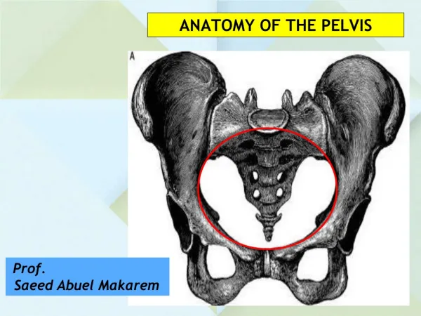
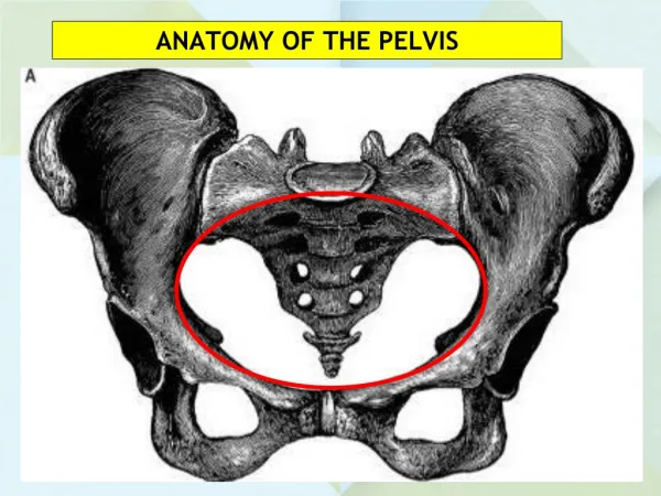
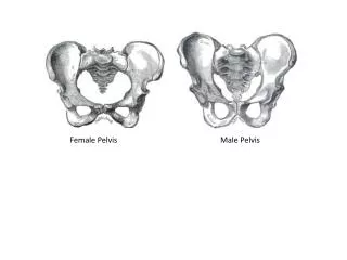
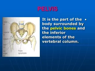
The Pelvis. The pelvis is the region of the trunk that lies below the abdomen; the abdominal and pelvic cavities are continuous.

E N D
The pelvis is the region of the trunk that lies below the abdomen; the abdominal and pelvic cavities are continuous.
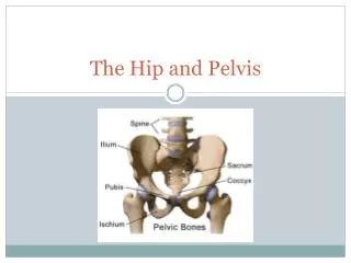
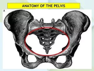
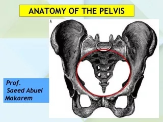
Surface AnatomyIliac Crest; Anterior Superior Iliac Spine; Posterior Superior Iliac Spine; Tubercle of Iliac Crest; Pubic Tubercle; Pubic Symphysis; Pubic Crest; Spinous Processes Sacrum; Sacral Hiatus; Coccyx.
Examine again the division of the lower part of the abdominal aorta in front of the fourth lumbar vertebra into two common iliac arteries. Follow the common iliac arteries to their termination in front of the sacroiliac joints. Trace the external iliac artery along the pelvic brim to the inguinal ligament.
Identify the inferior epigastric and deep circumflex iliac branches. Clean the origin of the internal iliac artery and follow it down into the pelvis, where it ends near the upper margin of the greater sciatic foramen by bifurcating into anterior and posterior divisions. Identify the median sacral artery.
Expose and clean the external, internal and common iliac veins.
Identify and clean the right and left ureters as they enter the pelvis in front of the bifurcation of the common iliac arteries.
Examine again the lumbaosacral trunks and the obturator nerves as they enter the pelvis. Do not disturb them in the pelvis at the present time.
Examine again the lower ends of the lumbar sympathetic trunks and ganglia. Demonstrate the continuity of the trunks with those in the pelvis.
Trace the hypogastric plexus over the sacral promontory,but do not disturb it within the pelvis at this time.
Trace the testicular arteries to the deep inguinal rings, or the ovarian arteries to the pelvic brim.
Examine again the pelvic colon and identify the pelvic mesocolon. Follow the superior rectal artery to the pelvic inlet and to the posterior surface of the rectum.
Examine again the femoral nerve and the lateral femoral cutaneous nerve. Trace the femoral nerve as it emerges from the lateral border of the psoas muscle, downward and laterally in the groove between the psoas and the iliacus.
Note that it lies under the cover of the fascia iliaca and passes behind the inguinal ligament to enter the thigh lateral to the femoral artery. Follow the lateral cutaneous nerve of the thigh across the iliacus muscle and verify that it enters the thigh behind the lateral end of the inguinal ligament.
Peritoneum. Examine the peritoneum and trace it down the anterior and lateral surfaces of the rectum. Follow the peritoneal reflection from the rectum onto the upper part of the posterior surface of the vagina, which forms the rectouterine pouch (pouch of Douglas).
The peritoneal recess on each side of the rectum is referred to as the pararectal fossa. Note that the pararectal fossa and the rectouterine pouch are continuous with one another.
Trace the peritoneum over the posterior surface of the uterus, over the fundus, and down over the anterior surface of the uterus. It then passes onto the posterior surface of the bladder for a short distance, thus forming the uterovesical pouch.
Trace the peritoneum over the upper surface of the bladder and then forward onto the anterior abdominal wall. Note that the outline of the empty bladder is not easily seen. Note also that the peritoneum runs from the superior surface of the bladder to the lateral walls of the pelvis.
Observe that the normal uterus is bent forward so that its anterior surface faces forward and inferiorly and overhangs the bladder (anteverted and anteflexed).
Note that the peritoneum almost completely covers the uterus and extends laterally on each side of the uterus as a two-layered fold of peritoneum to the side wall of pelvis. This fold is known as the broad ligament. Identify in the broad ligament the following structures:
1.The uterine tube. This lies in the free border of the broad ligament.It opens into the peritoneal cavity by means of its funnel-shaped lateral extremity, the infundibulum. The free edge of the funnel is broken up into a number of finger-like processes known as the fimbriae,which are draped over the ovary.
2. The ovary is a small oval body attached to the back of the broad ligament by the mesovarium.
3. The mesosalpinx is that portion of the broad ligament which lies between the mesovarium and the uterine tube.
4. The suspensory ligament of the ovary is that part of the broad ligament which lies lateral to attachment of the mesovarium; it contains the ovarian vessels and nerves.
5. The round ligament of the ovary produces a ridge of peritoneum on the posterior surface of the broad ligament.
6. The round ligament of the uterus produces a ridge of peritoneum on the anterior surface of the broad ligament that can be trace from the lateral edge of the uterus to the deep inguinal ring.
With the fingers, carefully mobilize the rectum from the anterior surface of the sacrum. Dissect off the peritoneum and pelvic fascia from the posterior and lateral wall of the pelvis.
Clean the rectum and observe that the lower third is completely devoid of peritoneum, since it lies below the lowest part of the rectouterine pouch. Note that the rectum expands just above the pelvic floor to form the ampulla of the rectum. Once the rectum passes through the pelvic floor, it becomes the anal canal.
Follow the superior rectal artery downward along the posterior surface of the rectum. Note that it divides into two branches that pass down on either side of the rectum and anastomose with the right and left middle rectal arteries, which are branches of the internal iliac artery.
Identify the hypogastric and the left pelvic plexuses. The hypogastric plexus descends into the pelvis and divides into right and left pelvic plexuses. The fine nerve threads of the pelvic plexus will be found in the fascia surrounding the internal iliac artery.
Trace the pelvic part of the sympathetic trunk inferiorly. Note that above and posterior to the common iliac vessels it is continuous with the abdominal part of the trunk.
Below it descends posterior to the rectum and medial to the anterior sacral foramina. It has four or five segmentally arranged ganglia. The trunks end below by uniting in the midline anterior to the coccyx in the ganglia impar. Identify but do not disturb the sacral plexus.
Ureter. Identify and clean the left ureter. Verify that it enters the pelvis by crossing the bifurcation of the common iliac artery in front of the sacroiliac joint. It runs downward and backward in front of the internal iliac artery and behind the ovary.
Follow the ureter to the region of the ischial spine and trace it forward, beneath the base of the broad ligament. Note that the ureter is crossed superiorly from lateral to medially by the left uterine artery at this point. Note also that the ureter lies lateral to the cervix and the left lateral fornix of the vagina.
Now trace the ureter forward across the pelvic floor to the bladder. Insert a fine probe into the ureteric orifice within the bladder and verify its oblique course through the bladder wall.
Clean and study the internal iliac artery and its branches. Note that it arises from the common iliac artery and passes down into the pelvis medial to the external iliac vein and anterior to the internal iliac vein. The main branches are visceral and parietal and are as follows:
Visceral Branches1. The umbilical artery gives origin to the superior vesical artery. The obliterated remnant then ascends to the umbilicus as the lateral umbilical ligament.
2. The inferior vesical artery runs forward to the base of the bladder, which it supplies and in addition gives branches to the ureter.
4. The uterine artery runs forward and medially to enter the base of the broad ligament, where it ascends along the lateral margin of the body of the uterus. Note its relationship to the ureter.
Parietal Branches1.The iliolumbar artery runs upward and laterally, deep to the common iliac vessels, and terminates by supplying the iliacus, psoas, and quadratus lumborum muscles.
2. The lateral sacral artery or arteries ( there are usually two ) pass medially to descend in front of the anterior sacral foramina.
3. The obturator artery runs anteriorly with the obturator vein and nerve to reach the obturator canal, where it enters the adductor compartment of the thigh.
4.The internal pudendal artery leaves the pelvis between the piriformis and coccygeus muscles, through the lower part of the greater sciatic foramen. It will be dissected in the gluteal region and again in the perineum.