Download
1 / 20
230 likes | 565 Views
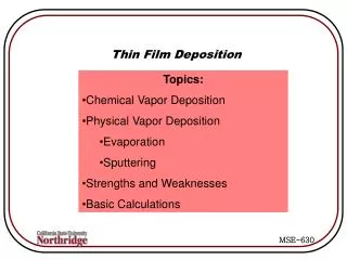
Thin Film Cyclic Voltammetry. Equipment for film voltammetry. potentiostat. electrode material. insulator. reference. Electroactive film. N 2 inlet. counter. working electrode. E-t waveform. Cyclic voltammetry. E, V. Electrochemical cell. time.

E N D
Equipment forfilm voltammetry potentiostat electrode material insulator reference Electroactive film N2 inlet counter working electrode E-t waveform Cyclic voltammetry E, V Electrochemical cell time
Ideal, reversible thin layer cyclic voltammogram Example cobalt complex: LCoIII+ e- LCoII Q = nFAGTGT = total surface concentration of electroactive species A = electrode area, F = Faraday’s constant Ep Ip reversible peak current Ip increases linearly as scan rate () is increased; And DEp = 0. Rate constants can be obtained by increasing to drive the CV into a kinetically limited situation where DEp > 0. Q = area under reduction curve
Many types of electroactive films Ferrocene SAM Electroactive polymer Protein SAM SAM = self assembled monolayer
Real CVs, include Charging current And some non-ideality
Electrochemistry of proteins in solution • electrode fouling, proteins denature • large size means small D, tiny signals • need lots of protein Thin Film Electrochemistry of Proteins Protein (monolayer) electrode Measure current Apply voltage Information obtained: Redox potentials, free energies, re-organization energies 2. Redox mechanism: protonation/deprotonation and chemical reaction steps Kinetics and thermodynamics of catalytic reactions 4. Biosensors
One way to make a stable protein film A lipid-protein film enzyme Electrode • Many other types of films possible - polyions, Adsorbed, crosslinked, etc.
Forward peak Reversible Peaks for Direct electron Transfer; Peak shapes, sizes, and Ep reveal details of redox chemistry Nearly ideal Reversible ET Reduction Of FeIII Oxidation Of FeII Reverse peak
Kinetically limited CV at 0.1 V s-1 for 40nm myoglobin (Mb)-polyion film on a PG electrode in pH 5.5 buffer at 35 oC. Example where rate constants can be obtained by increasing to drive the CV into a kinetically limited situation; DEp >> 0. Mb is another iron heme protein, peaks are for redox reactions of iron. Value of ks (s-1)cas be obtained by fitting data to theoretical curves of DEpvs. log scan rate or by fitting with best fit digital simulations of the CVs.
Prof. John Schenkman, Pharmacology, Cell Biology, Uconn Health Center Human Metabolic Enzymes: CytP450s in LbL polyion films: • ET reduction rates from CV depend on spin state of cyt P450 iron heme (low spin fastest); conformational equilibria • rates of oxidation by peroxide depend on spin state (high spin fastest) and secondary structure
Thin Film voltammetry of human cyt P450s LbL films of cyt P450s and polyions on pyrolytic graphite electrodes. Polyions are purple strands and proteins are green/red ribbons . Thickness 10-25 nm Sadagopan Krishnan,AmilaAbeykoon,John B. Schenkman, and James F. Rusling, Control of Electrochemical and Ferryloxy Formation Kinetics of Cyt P450s in Polyion Films by Heme Iron Spin State and Secondary Structure, J. Am. Chem. Soc. 2009, 131, 16215–16224.
Spectral characterization of cyt P450 films PFeIII PFeIII PFeII-CO UV-vis spectra of cyt P450 films on aminosilane-functionalized fused silica slides: (A) CO difference spectrum confirming native proteinin PEI(/PSS/cyt P450 1A2)6 film after reducing to the ferrous form and purging the pH 7 buffer with CO; (B) ferric high spin form of enzyme in PEI(/PSS/cyt P450 1A2)6;and (C) ferric low spin form of enzyme in PSS(/PEI/cyt P450cam)6 film.
Cyclic Voltammetry and rate constant (ks) estimates Assuming simple electron transfer model Background subtracted cyclic voltammograms of LbL films on PG electrodes in anaerobic 50 buffer + 0.1 M NaCl, pH 7.0 P450 2E1 P450 cam Rate const. estimation for cyt P450/polyion films experimental () peak separation (Ep) corrected for scan rate independent non-kinetic contribution. Lines for Butler-Volmer theory for the rate constant (ks) shown and a= 0.5.
The simple reversible theory did not fit peak potential vs. scan rate data, so complex model Lines were from digital simulation using
Conclusions for cyt P450 ET from thin Film voltammetry: • low spin cyt P450cam, ks = 95 s-1 mixed spin cyt P450 1E2, ks = 18 s-1 (80% high spin) high spin cyt P450 1A2, ks = 2.3 s-1 • ks for the reduction step correlates with spin state of the iron heme in the cyt P450, as found for solution reductions • rates of oxidation by peroxide depend on spin state (high spin fastest) also
Divided cell – keep products apart Undivided cell – sacrificial anode can be used e.g. Cu Cu2+ + 2e
Divided Electrolysis Cell for synthetic use Counter electrode Large working electrode + ref