Download
1 / 40
400 likes | 514 Views
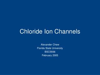
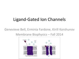
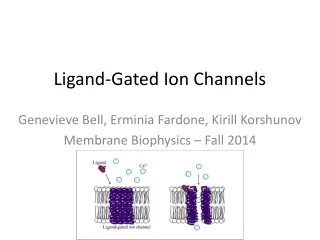
Regulation of ion channels by lipid signaling. Hana Cho Department of Physiology, Sungkyunkwan University School of Medicine. acetylcholine. GIRK. OUT. M 2. M 2. bg. a. bg. IN. a. GTP. K +. GDP. GIRK channels in atrial myocytes. acetylcholine. ACh. ACh.

E N D
Regulation of ion channels by lipid signaling Hana Cho Department of Physiology, Sungkyunkwan University School of Medicine
acetylcholine GIRK OUT M2 M2 bg a bg IN a GTP K+ GDP GIRK channels in atrial myocytes acetylcholine
ACh ACh a b c d GIRK channels are regulated by membrane phospholipid, PIP2 500 pA 2 min Logothetis & Zhang. J. Physiol. 520:630, 1999
PIP2: a substrate of phospholipase C DAG PIP2 IP3
a DAG PLC Gq IP3 Is PIP2 a signaling molecule? GIRK PIP2
Cardiac GIRK channels: Inhibited by GqPCRs via PIP2 depletion J Biol Chem 276:159 in a receptor-specific manner ; J Biol Chem 277: 27742; PNAS 102: 4643 ACh ACh ACh ET-1 BK PE
Excitable cells endocrine cells neurons myocytes Electrical signals underly functions of excitable cells
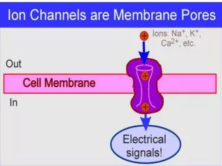
Ion channels/transporters are basis of electrical signals Ion current through ion channels electrical signal
Electrical signals are measured by patch clamp technique and microfluorometry (Fura-2)
Nobel Prize in Chemistry, 2008 The Nobel Prize in Chemistry in 2008 awarded Osamu Shimomura, Martin Chalfie and Roger Y. Tsien "for the discovery and development of the green fluorescent protein, GFP". Osamu Shimomura Martin Chalfie Fura-2 Roger Tsien
objective 505 nm Ca2+ signals can be studied using Fura-2 Ca2+ 340 & 380 nm PC AD/DA converter Xe & Monochromator Photodiode
Ca2+ is a common second messenger for cellular output Ca2+ Shaping Ca2+ signals Cellular outputs contraction secretion
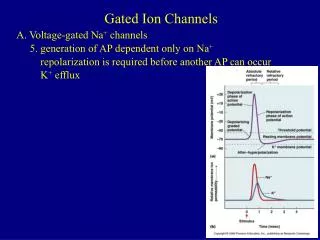
Patch clamp? Patch; a small piece of cell membrane Patch Clamp; imposing on a membrane patch a defined voltage (" volt age-clamp") with the purpose to measure the resulting current for the calculation of the patch conductance voltage- clamp of a membrane patch. Neher and Sakmann in 1976 Nobel prize; 1992
Voltageclamp technique Current to voltage converter
When the pipette touches the cell membrane and forms a high resistance seal (~1GOhm), you are in the "cell attached" recording configuration.
Recording equipment • Patch-clamp set의 일반적 구성은 다음과 같다 • Optics; Microscope, TV camera & monitor • 2. Mechanics; Vibration isolation table, Micromanipulator • 3. Electronics; Patch-amplifier,, ADDA converter Computer • 4. Environment; bath • 5. 기타; Feraday cage, Photometry system
Overview of the recording set-up: microscope, Micromanipulator, patch clamp amplifier, computer.
A close-up view of the recording scope. The recording electrode is attached to the blue headstage.
A close-up view of the microscope stage. The cells are plated on a glass coverslip that is adhered to the bottom of the plastic dish. The electrode holder does not have a pipette on it in this picture.
Patch clamper is using the computer to run the patch clamp amplifier and collect data.
The top trace is the current response of the electrode in the bath. The bottom trace shows the small voltage step that is being applied repeatedly.
As the seal begins to form, the horizontal lines of the current trace move closer together (compare to previous photo).
Patch clamper is gently sucking on the back of the syringe that is connected to the back of the pipette. This suction will help seal the tip of the pipette onto the cell.
The amplifier is used to compensate, i.e. to electronically remove, the pipette capacitance from the current recording.
After electrically removing the pipette capacitance, the vertical spikes are gone.
To break into the whole cell recording mode, additional suction is applied to the syringe. As shown here, you'll know when a whole cell recording in acheived because the capacitative transients (the vertical "spikes" due to the cell capacitance) are large and have a slow decay.
GIRK a M2 DAG PLC PIP2 Gq Gi/o IP3 Cardiac GIRK channels: Inhibited by GqPCRs via PIP2 depletion J Biol Chem 276:159 in a receptor-specific manner ; J Biol Chem 277: 27742; PNAS 102: 4643 ACh ACh ACh ET-1 BK PE
Finally we can propose comprehensive model for GIRK channel regulation by Gq-coupled receptors
ET-1 ACh ACh ET-1 5 pA 20 sec 2 pA 20 sec Recording GIRK current in cell-attached patches
Fluorescence Recovery After Bleaching (FRAB) to measure diffusion of second messenger (PIP2) confocal image Slope = R0 [R 02 + 8Dt]-1/2 where R0 is the half-width (1/e2) of the bleach D=0.000390.000038 mm2/s (n=5)
Acknowledgement SKKU, SBRI Jin Young Yoon Sun Ok Choi Slee Lee Hyun Tae Cho SNU Dr. Won-Kyung Ho Dr. Suk-Ho Lee Jong Woo Shon Doyun Lee