Download
1 / 31
330 likes | 553 Views
SPINAL CORD INJURY. Case in Neurosurgery Section A USTFMS.

E N D
SPINAL CORD INJURY Case in Neurosurgery Section A USTFMS
A 50 y/o male was thrown from his motorcycle and landed on his head. He was then brought to a hospital some 30 km far reclining just at the back seat of a car. He continues to complain of persistent nape pain that radiates down to the lateral aspect of his right forearm. On arrival at the ER, he was noted to have difficulty moving all extremities. He has numbness from neck down with no difficulty breathing.
Physical examinations revealed the following: • Diminished pain and temperature sensation from neck down • Position and vibratory senses intact
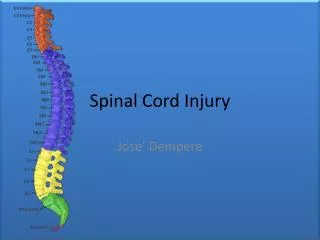
Spinal Cord Injury • an insult to the spinal cord resulting in a change, either temporary or permanent, in its normal motor, sensory, or autonomic function • International Standards for Neurological and Functional Classification of Spinal Cord Injury - a widely accepted system describing the level and the extent of injury based on a systematic motor and sensory examination of neurologic function
The following terminology has developed around classification of SCI: • Tetraplegia (quadriplegia) - injury to the spinal cord in the cervical region ; loss of muscle strength in all 4 extremities • Paraplegia - injury in the spinal cord in the thoracic, lumbar, or sacral segments, including the cauda equina and conus medullaris
SCI can be sustained through: • Destruction from direct trauma • Compression by bone fragments, hematoma, or disk material • Ischemia from damage or impingement on the spinal arteries
Spinal Cord Injuries • Complete or Partial 1) Complete = cause permanent quadriplegia or paraplegia, depending on the level of injury • Complete loss of motor function & sensation 2 or more levels below the bony injury
2) Partial: • Central cord syndrome – usually in elderly who suffer hyperextension injuries; motor, pain & temp preserved in LE but diminished in UE • Some functional recovery but seldom return to normal • Anterior cord syndrome – diminished motor function & pain, temp. sensation below level of injury; position, vibratory sensation & crude touch maintained • Prognosis for recovery- poor • Brown Sequard syndrome- result of penetrating injury; R or L half of sp.cord is transected; ipsilateral loss of motor fxn, proprioception, vibratory sensation; pain & temp. sensation lost on contraleteral side
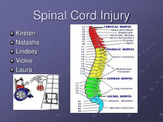
Key muscles = level of injury • C5 - Elbow flexors (biceps, brachialis) • C6 - Wrist extensors (brachioradialis, extensor carpi radialis longus and brevis) • C7 - Elbow extensors (triceps) • C8 - Finger flexors (flexor digitorum profundus) to the middle finger • T1 - Small finger abductors (abductor digiti minimi) • L2 - Hip flexors (iliopsoas) • L3 - Knee extensors (quadriceps) • L4 - Ankle dorsiflexors (tibialis anterior) • L5 - Long toe extensors (extensors hallucis longus) • S1 - Ankle plantar flexors (gastrocnemius, soleus)
Myotome • Each muscle in the body is supplied by a particular level or segment of the spinal cord and by its corresponding spinal nerve. The muscle, and its nerve make up a myotome.
C5 C6 C7 L2 L2 L3 L3
Types of SC Injury Trauma: crashes, falls, gunshots, diving accidents, war injuries, etc. Tumors Ischemia resulting from occlusion of spinal blood vessels, including dissecting aortic aneurysms, emboli, arteriosclerosis. Developmental disorders: spina bifida, meningomyelocoele, etc. Neurodegenerative diseases: spinocerebellar ataxia, etc. Demyelinative diseases: MS Transverse myelitis resulting from stroke, inflammation, etc. Vascular malformations: AVM, AVF, spinal hemangiona, cavernous angioma and aneurysm.
American Spinal Injury Association (ASIA) Classification • Based on neurological responses, touch and pinprick sensations tested in each dermatome, and strength of ten key muscles on each side of the body, • Traumatic spinal cord injury: 5 categories A: "complete" SC injury where no motor/sensory function is preserved in the sacral segments S4-S5. B: "incomplete" SC injury where sensory but not motor function is preserved below the neurological level and includes the sacral segments S4-S5 C: "incomplete" SC injury where motor function is preserved below the neurological level and more than half of key muscles below the neurological level have a muscle grade of less than 3, which indicates active movement with full range of motion against gravity. D: "incomplete" SC injury where motor function is preserved below the neurological level and at least half of the key muscles below the neurological level have a muscle grade of 3 or more. E: "normal" where motor/sensory scores are normal.
Anterior Cord Syndrome • Results from injury to the anterior part of the spinal cord, causing weakness and loss of pain and thermal sensations below the injury site but preservation of proprioception that is usually carried in the posterior part of the spinal cord
The Patient Partial Spinal Cord Traumatic Injury • Anterior Cord Syndrome • (C6 and below affected) • diminished motor function & pain, temp. sensation below level of injury; position, vibratory sensation & crude touch maintained
On-Site Diagnosis of Spinal Injury • Assume Spinal Cord Injury if: • Evidence of a head injury with ongoing change in the person's level of consciousness. • Complaints of severe pain in head or back. • A substantial force was on sustained on back or head. • Complaints of weakness, numbness or paralysis or lacks control of his or her limbs, bladder or bowel. • Neck or back is twisted or positioned oddly.
On-Site Diagnosis of Spinal Injury • Questions to ask: • name • location of injury • time of day • Incident • If the person cannot accurately answer at least the first three questions, he or she is not a "reliable patient," and a spinal cord injury cannot be ruled out.
On-Site Diagnosis of Spinal Injury • Look for any signs of drug or alcohol use. • the patient is not reliable if (+). • Check for "distracting injuries" • injuries painful enough to distract the injured patient due to severe pain at the spine and head. Spinal cord injury cannot be ruled out.
On-Site Diagnosis of Spinal Injury • Instruct the injured person to move his or her fingers and toes. • If any motion is difficult or impossible, or if the person is abnormally weak, there may be a spinal injury. • Squeeze the fingers and toes gently. • If any numbness or tingling, the spine may be injured.
On-Site Diagnosis of Spinal Injury • Palpate each vertebrae with moderate pressure, from the top of the neck to the bottom of the back. Look for any signs of pain.
While waiting for paramedics, patient’s neck should always be immobilized • Rigid cervical collar, skull tract, halo brace etc.
ER Diagnosis of Spinal Injury • Detailed History • Details of event • Previous history of injuries • Physical Examination • Neurological Exam with more detailed attention.
ER Diagnosis of Spinal Injury • Neurological Exam • Mental status • Cranial nerves • Motor Strength • Sensation • Coordination • Reflexes • Gait
Diagnosis • To diagnose the presence of spinal cord injuries, these imaging modalities may be used: • Plain film (X-ray) • CT Scan • MRI
Plain Film • Advantages: • Rapid survey of the bony spine • Presence, level and type of lesion • Fracture • Dislocation • Subluxation • Disadvantages: • Cannot adequately evaluate lower cervical and upper thoracic spine because of the shoulder girdle • A single view is inadequate: • 3 views at least
CT Scan • Advantage: • Good for Characterizing fractures on Plain Film • Characterize suspicious lesions on Plain Film • Can Visualize C7-T1 when not seen on Plain Film • Combination of Plain Film and CT: false negative of less than .1% • Especially useful in posterior vertebral injuries • Fine Slice with Sagittal and Coronal Reconstruction • Must include the entire vertebral body above and below the lesion of interest
MRI • Advantages: • Best in demonstrating injuries to soft tissue • the neural elements, especially the spinal cord • Contusions • Ischemia • Traumatic disk herniations • Ligamentous injuries • Canal compromise • Vertebral Artery injury • If the neurologic level of injury does not match the area of injury identified by standard radiographs
Prevention • Safety tips for preventing the occurrence of a SCIMotor vehicles are the leading cause of SCI in the United States for people under age 65 (Berkowitz 1998). Here are some safety tips for driving and riding in motor vehicles. • Always wear a seat belt. • Secure or buckle children into age- and weight-appropriate child safety seats. • Secure or buckled children under 12 years old in the back seat to avoid air bag injuries. • Never drive under the influence of alcohol or drugs. • Do not ride in a car with a driver who is impaired by alcohol or drugs.. • Prevent others from driving while impaired by alcohol or drugs. http://www.cdc.gov/ncipc/factsheets/sciprevention.htm
Prevention • The following might have prevented spinal cord injury to our patient: • Avoid riding motorcycles. • Always wear a helmet. • Proper transport of the injured person. (Stabilize neck and back, and avoid unnecessary changes in position)