Download
1 / 15
200 likes | 256 Views
THE PAROTID REGION. Dr. Ahmed Fathalla Ibrahim. THE PAROTID REGION. It includes: The parotid salivary gland The structures related to the gland. PAROTID GLAND. THE PAROTID GLAND. DEFINITION: It is the largest of the salivary glands

E N D
THE PAROTID REGION Dr. Ahmed Fathalla Ibrahim
THE PAROTID REGION • It includes: • The parotid salivary gland • The structures related to the gland
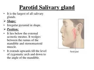
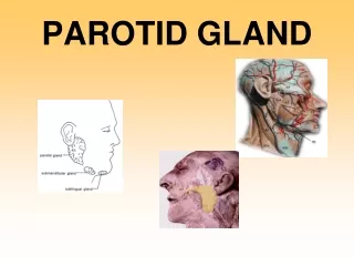
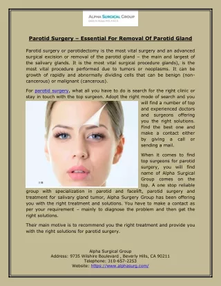
THE PAROTID GLAND • DEFINITION: It is the largest of the salivary glands • SITE: It lies below the auricle, occupying the region between ramus of mandible & mastoid process • EXTENT: • Superiorly: to zygomatic arch • Inferiorly: to angle of mandible • Anteriorly: to overlap posterior border of masseter • Posteriorly: to overlap anterior border of sternomastoid • SHAPE: Pyramidal
THE PAROTID GLAND • SUBDIVISIONS: • Main gland • Accessory gland: above parotid duct • CAPSULE: • Derived from deep fascia of neck (cervical fascia) • Its superficial layer is attached to zygomatic arch & extends to cover masseter • Its deep layer is attached to mandible, styloid & mastoid processes • A thickening of deep fascia extends from styloid process to angle of mandible (stylomandibular ligament) & separates the capsule of parotid from that of submandibular gland • It is tense (swellings of parotid gland are painful)
THE PAROTID GLAND • RELATIONS: • Superficial: skin, superficial fascia, great auricular nerve, superficial parotid (preauricular) lymph nodes • Anteromedial: posterior border of ramus of mandible + muscles attached to ramus (masseter, medial pteygoid) • Posteromedial:mastoid process + muscles attached to it (sternomastoid, posterior belly of digastric), styloid process + muscles attached to it (stylohyoid, styloglossus, stylopharyngeus), carotid sheath & its contents (internal jugular vein, internal carotid artery, 9th, 10th, 11th & 12th cranial nerves) • Medial: pharyngeal wall
STRUCTURES WITHIN THE PAROTID GLAND • Termination of facial nerve & beginning of its five terminal motor branches : most superficial structures • Terminations of superficial temporal & maxillary veins + the whole retromandibular vein + beginning of its two divisions (anterior & posterior) • Termination of external carotid artery & beginning of its two terminal branches (superficial temporal & maxillary): deepest structures • Deep parotid lymph nodes: embedded within substance of the gland
PAROTID DUCT • LENGTH: Two inches • COURSE & RELATIONS: • Emerges from anterior border of gland • Runs obliquely forwards, superficial to masseter & below transverse facial artery & accessory parotid • TERMINATION: • Pierces: buccal pad of fat, buccopharyngeal fascia, buccinator muscle & buccal mucosa • Opens: into the vestibule of mouth, opposite the crown of upper 2nd molar tooth • APPLIED ANATOMY: The oblique passage of the duct act as a valve-like mechanism & prevents inflation of the duct during blowing • SURFACE ANATOMY: It is represented by the middle 1/3 of a line extending from the tragus of the auricle to a point midway between the ala of nose & upper lip
NERVE SUPPLY • PARASYMPATHETIC (SECRETORY): • Origin: inferior salivary nucleus (medulla) • Preganglionic fibers: run along the lesser petrosal nerve (branch of tympanic of glossopharyngeal (9th cranial) • Ganglion: fibers relay in the otic ganglion (infratemporal fossa) • Postganglionic fibers: reach the parotid gland along auriculotemporal nerve (branch of mandibular of trigeminal) • SYMPATHETIC:Postganglionic sympathetic fibers reach the gland as a plexus around external carotid artery
BLOOD SUPPLY • ARTERIES: External carotid • VEINS: Retromandibular vein
LYMPHATIC DRAINAGE • Into superficial & deep parotid lymph nodes • Finally into deep cervical lymph nodes