Download

1 / 81
830 likes | 882 Views
The Gluteal Region. Dr. Fadel Naim Orthopedic Surgeon Faculty of Medicine IUG. The lower limbs specialized for locomotion The primary function of the lower limbs: Support the weight of the body Provide a stable foundation in: Standing Walking Running

E N D
The Gluteal Region Dr. Fadel Naim Orthopedic Surgeon Faculty of Medicine IUG
The lower limbs specialized for locomotion The primary function of the lower limbs: Support the weight of the body Provide a stable foundation in: Standing Walking Running Similar in structure in many respects to the upper limbs Have less freedom of movement The upper limb is united to the trunk by only a small joint, (the sternoclavicular joint) The two hip bones articulate: Posteriorly with the trunk at the strong sacroiliac joints Anteriorly with each other at the symphysis pubis. The lower limbs are more stable
Organization Of The Lower Limb • The lower limbs are divided into different regions and compartments • The regions: • The gluteal region • The thigh • The knee • The leg • The ankle • The foot • The thigh and the leg are compartmentalized • Each compartment with own muscles • Perform group functions • Own distinct nerve and blood supply
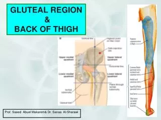
The Gluteal Region • The gluteal region bounded • superiorly by the iliac crest • inferiorly by the fold of the buttock. • The region is largely made up of the gluteal muscles and a thick layer of superficial fascia
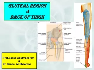
A.Level of Iliac crest (L4,) B. intergluteal cleft C. buttock D. gluteal fold E. thigh F. Gluteal sulcus
The gluteal region contains: • Bones • Ligaments • Muscles • Vessels • Nerves
Dermatomes Cutaneous Nerve supply: • Upper lateral quadrant: Lateral branches of iliohypogastric (L1) and T12 • Upper medial quadrant: Posterior rami of L1,2,3 & S1,2,3 • Lower lateral quadrant: branches from lateral cutaneous nerve of thigh (L2,3) • Lower medial quadrant: branches from posterior cutaneous nerve of thigh (S1,2,3) • Skin in the floor of the intergluteal cleft: branches from lower sacral and coccygeal nerves
Fascia Of The Buttock • The superficial fascia is thick and impregnated with large quantities of fat. • It contributes to the prominence of the buttock. • The deep fascia is continuous below with the deep fascia of the thigh (fascia lata). • It is attached to the iliac crest. • In the gluteal region, it splits to enclose the gluteus maximus muscle • It continues as a single layer that covers the outer surface of the gluteus medius
TheIliotibialTract • On the lateral surface of the thigh, thickened to form a strong, wide band • From the tubercle of the iliac crest and below to the lateral condyle of the tibia • Forms a sheath for the tensor fasciae latae muscle • Receives the greater part of the insertion of the gluteus maximus
Bones Of The Gluteal Region Hip bone • The ilium • The ischium • The pubis • Meet one another at the acetabulum • Articulate with the sacrum at the sacroiliac joints • Form the anterolateral walls of the pelvis • Articulate with one another anteriorly at the symphysis pubis.
The upper flattened part of the hip bone • The iliac crest • Can be felt through the skin along its entire length • Anterior superior iliac spine • Anterior inferior iliac spine • Posterior superior iliac spine • posterior inferior iliac spine. • The iliac tubercle lies about 5 cm behind the anterior superior spine. • Greater sciatic notch The Ilium
Theischium • L shaped • The body • An upper thicker part • The ramus • A lower thinner part, • The ischial spine • Projects from the posterior border of the ischium and intervenes between the greater and lesser sciatic notches. • Converted into greater and lesser sciatic foramina by the presence of the sacrospinous and sacrotuberous ligaments • The ischial tuberosity • Forms the posterior aspect of the lower part of the body of the bone.
Thepubis • Divided into: • A body • A superior ramus • An inferior ramus • The bodies of the two pubic bones articulate with each other in the midline anteriorly at the symphysis pubis • The superior ramus joins the ilium and ischium at the acetabulum • The inferior ramus joins the ischial ramus below the obturator foramen. • The obturator foramen in life is filled in by the obturator membrane • The pubic crest forms the upper border of the body of the pubis, and it ends laterally as the pubic tubercle
TheAcetabulum • On the outer surface of the hip bone is a deep depression, called the acetabulum • Articulates with the head of the femur to form the hip joint • The inferior margin of the acetabulum is deficient and is marked by the acetabularnotch • The articular surface of the acetabulum is limited to a horseshoe shaped area and is covered with hyaline cartilage. • The floor of the acetabulum is non-articular and is called the acetabular fossa
Femur • Articulates above with the acetabulum and below with the tibia and the patella • The upper end of the femur has • A head • A neck • Greater and lesser trochanters • Thehead forms about two thirds of a sphere • Articulates with the acetabulum of the hip bone to form the hip joint
Femur Fovea capitis • A small depression in the center of the head for the attachment of the ligament of head. • Part of the blood supply to the head of the femur from the obturator artery is conveyed along this ligament and enters the bone at the fovea
The Neck of the Femur • Connects the head to the shaft • Pass downward, backward, and laterally • Makes an angle about 1250 with the long axis of the shaft. • (Slightly less in the female) • The size of this angle can be altered by disease
The Greater And Lesser Trochanters • Large eminence situated at the junction of the neck and the shaft • Connecting the two trochanters are the intertrochanteric line anteriorly, where the iliofemoral ligament is attached • Prominent intertrochanteric crest posteriorly, on which is the quadrate tubercle
Linea Aspera • The shaft of the femur is smooth and rounded on its anterior surface but posteriorly has a ridge, the linea aspera • Attachment of muscles and intermuscular septa. • The margins of the linea aspera diverge above and below. • On the posterior surface of the shaft below the greater trochanter is the gluteal tuberosity for the attachment of the gluteus maximus muscle
The medial margin continues below as the medial supracondylar ridge to the adductor tubercle on the medial condyle • The lateral margin becomes continuous below with the lateral supracondylar ridge. • The shaft becomes broader toward its distal end and forms a flat, triangular area on its posterior surface called the popliteal surface
The lower end of the femur has lateral and medial condyles, separated posteriorly by the intercondylar notch. • The anterior surfaces of the condyles are joined by an articular surface for the patella. • The two condyles take part in the formation of the knee joint. • Above the condyles are the medial and lateral epicondyles • The adductor tubercle is continuous with the medial epicondyle.
Arthritis Of The Hip Joint • The head of the femur can be palpated on the anterior aspect of the thigh just inferior to the inguinal ligament and just lateral to the pulsating femoral artery. • Tenderness over the head of the femur usually indicates the presence of arthritis of the hip joint.
Blood Supply ToThe Femoral Head • In the young, the epiphysis of the head is supplied by a small branch of the obturator artery, which passes to the head along the ligament of the femoral head. • The upper part of the neck of the femur receives a profuse blood supply from the medial femoral circumflex artery. • These branches pierce the capsule and ascend the neck deep to the synovial membrane. • As long as the epiphyseal cartilage remains, no communication • In adult anastomosis is present • Fractures of the femoral neck interfere with or completely interrupt the blood supply from the root of the femoral neck to the femoral head. • The blood flow along the small artery may be insufficient to sustain the viability of the femoral head • ischemic necrosis gradually takes place.
Coxa Valga And Coxa Vara • The neck of the femur is inclined at an angle with the shaft; the angle is about 1600 in the young child and about 1250 in the adult. • An increase in this angle is referred to as coxa valga • ( congenital dislocation of the hip) • In this condition, adduction of the hip joint is limited. • A decrease in this angle is referred to as coxa vara, • ( fractures of the neck of the femur and in slipping of the femoral epiphysis) • In this condition, abduction of the hip joint is limited. • Shenton's line is a useful means of assessing the angle of the femoral neck on a radiograph of the hip region
Subcapital Fracture • Fractures of the neck of the femur are common • The subcapital fracture • occurs in the elderly (common in women after menopause ) • usually produced by a minor trip or stumble. • Avascular necrosis of the head is a common complication. • If the fragments are not impacted, considerable displacement occurs. • The strong muscles of the thigh including the rectus femoris, the adductor muscles, and the hamstring muscles, pull the distal fragment upward, so that the leg is shortened • The gluteus maximus, the piriformis, the obturator internus, the gemelli, and the quadratus femoris rotate the distal fragment laterally, as seen by the toes pointing laterally.
Trochanteric fractures • In the young and middle aged as a result of direct trauma. • Extracapsular • Both fragments have a profuse blood supply. • If not impacted, the pull of the strong muscles will produce shortening and lateral rotation of the leg
Fractures of the shaft of the femur • usually occur in young and healthy persons. • In fractures of the upper third of the shaft of the femur • the proximal fragment is: • flexed by the iliopsoas • abducted by the gluteus medius and minimus • laterally rotated by the gluteus maximus, the piriformis, the obturator internus, the gemelli, and the quadratus femoris • The lower fragment is: • adducted by the adductor muscles • pulled upward by the hamstrings and quadriceps • laterally rotated by the adductors and the weight of the foot
Fractures Of The Middle Third Of The Shaft Of The Femur, • The distal fragment is pulled upward by the hamstrings and the quadriceps resulting in considerable shortening. • The distal fragment is also rotated backward by the pull of the two heads of the gastrocnemius
Fractures Of The Distal Third Of The Shaft • The distal fragment is smaller and is rotated backward by the gastrocnemius muscle to a greater degree • May exert pressure on the popliteal artery and interfere with the blood flow through the leg and foot • Considerable traction on the distal fragment is usually required to overcome the powerful muscles and restore the limb to its correct length before manipulation and operative therapy to bring the proximal and distal fragments into correct alignment.
Ligaments Of The Gluteal Region • The two important ligaments in the gluteal region : • Sacrotuberous and sacrospinous ligaments. • The function of these ligaments is to stabilize the sacrum and prevent its rotation at the sacroiliac joint by the weight of the vertebral column.
Ligaments Of The Gluteal Region • Sacrotuberous ligament • connects the back of the sacrum to the ischial tuberosity • Sacrospinous ligament • connects the back of the sacrum to the spine of the ischium
SI Ligaments: Sacrotuberous Ligament: Arises from ischial tuberosity to blend in with inferior fibers of posterior SI ligaments Sacrotuberous Ligament Ischial Tuberosity
SI Ligaments: Sacrospinous Ligament: Originates from the ischial spine and attaches to the coccyx Sacrospinous Ligament
Foramina Of The Gluteal Region • The two important foramina in the gluteal region are • The greater sciatic foramen • The lesser sciatic foramen
Greater Sciatic Foramen • Formed by the greater sciatic notch of the hip bone and the sacrotuberous and sacrospinous ligaments. • It provides an exit from the pelvis into the gluteal region. • The following structures exit the foramen • Piriformis • Sciatic nerve • Posterior cutaneous nerve of the thigh • Superior and inferior gluteal nerves • Nerves to the obturator internus and quadratus femoris • Pudendal nerve • Superior and inferior gluteal arteries and veins • Internal pudendalartery and vein
Structures passing through the greater sciatic foramen Above the piriformis: Superior gluteal vessels & nerve Piriformis: an important landmark Below the piriformis: Inferior gluteal vessels & nerve Sciatic nerve Posterior cutaneous nerve of thigh Pudendalnerve & Internal pudendal vessels Nerve to obturator internus Nerve to quadratus femoris
Lesser Sciatic Foramen • Formed by the lesser sciatic notch of the hip bone and the sacrotuberous ligaments. • Entrance into the perineum from the gluteal region. • Enables nerves and blood vessels that have left the pelvis through the greater sciatic foramen above the pelvic floor to enter the perineum below the pelvic floor. • The following structures pass through the foramen • Tendon of obturator internus muscle. • Nerve to obturator internus. • Pudendal nerve. • Internal pudendal artery and vein.
Structures passing through the lesser sciatic foramen Entering: Pudendal nerve & Internal pudendal vessels Exiting: Tendon of obturator internus Nerve to obturator internus
Gluteal Muscles • The gluteal muscles share a common compartment but are organized into two layers, superficial and deep: • Thesuperficial layerconsists of: • The three large glutei (maximus, medius, and minimus) • The tensor of the fascia lata. • All have proximal attachments to the posterolateral (external) surface and margins of the ala of the ilium • Mainly extensors, abductors, and rotators of the thigh.
Gluteal Muscles • Thedeep layerconsists of: • Smaller muscles covered by the inferior half of the gluteus maximus • Piriformis • Obturator internus • Gemelli • Quadratus femoris • All have distal attachments on or adjacent to the intertrochanteric crest of the femur. • Lateral rotators of the thigh • Stabilize the hip joint • Working with the strong ligaments of the hip joint to steady the femoral head in the acetabulum.
Muscles of the Gluteal Region • Gluteus maximus • Gluteus medius • Gluteus minimus • Tensor fascia lata • Piriformis • Superior Gemellus • Inferior Gemellus • Obturator internus • Quadratus femoris
Gluteus Maximus • The largest muscle in the body. • Superficial in the gluteal region • largely responsible for the prominence of the buttock. • Origin: • the outer surface of the ilium • the posterior surface of the sacrum and coccyx • the sacrotuberous ligament • Insertion: • The fibers pass downward and laterally • Most are inserted into the iliotibial tract • Some of the deeper fibers are inserted into the gluteal tuberosity of the femur. • Nerve supply: • Inferior gluteal nerve.