Download
1 / 79
790 likes | 803 Views
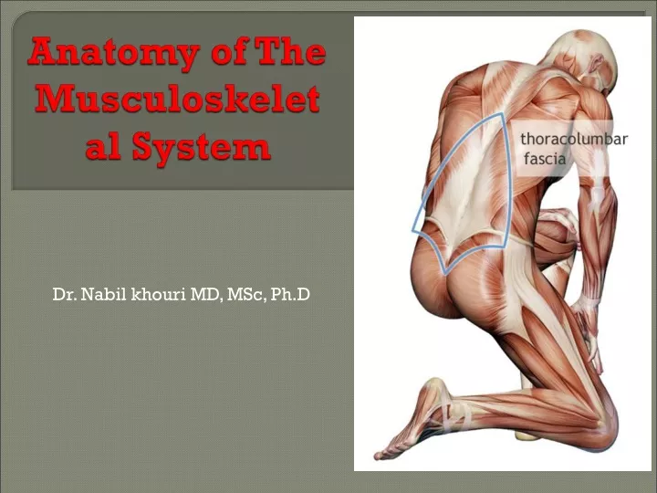
Anatomy of The Musculoskeletal System. Dr. Nabil khouri MD, MSc, Ph.D. What we will study! The Skeletal system “Objectives”. Skeletal system: is made of Bones that is a hard supporting tissue Bones are used to make up the skeleton Found in many forms including:

E N D
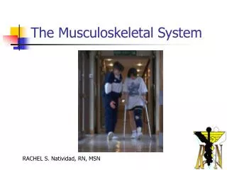
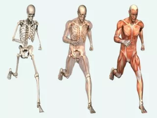
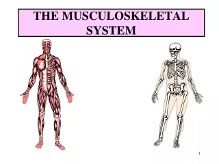
Anatomy of The Musculoskeletal System Dr. Nabil khouri MD, MSc, Ph.D
What we will study! The Skeletal system “Objectives” Skeletal system: is made of Bones that is a hard supporting tissue Bones are used to make up the skeleton Found in many forms including: “small, large, long, short and flat” Bones are held together by Joints which allow and/or restrict movements. Movements are performed by Muscle upon their contractions Muscle is made of muscular tissue
Objectives • Divisions of the Skeleton • Classification of Bones • Major bony landmarks
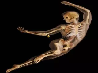
Bones: Forms In the skeleton and are arranged into Axial and appendicular groups • Vertebral Column 26 • Axial skeleton • Skull 22 • Hyoid bone 1 • Ribs and sternum 25 • ------- • Appendiclular skeleton • Upper Extremities 64 • Lower Extremities 62 • -------- • Auditory bones 6 • -------- • The total number of bones 206
Function of Bones • support (eg: pelvis, legs) • protect (eg: skull, vertebrae) • mineral storage (eg: calcium, phosphate, inorganic component) • movement (eg: walk, grasp objects) • blood-cell formation (eg: red bone marrow) • Cellular components include • Osteoblasts: secrete organic part of bone matrix = osteoid • Osteocytes: mature bone cells, maintain bone matrix
Divisions of the Skeleton • The Axial skeleton • The skull • The sternum • The ribs • The vertebral column • The appendicular skeleton • Upper extremities • Lower extremities • The shoulder girdle • The pelvic girdle
Classification of Bones Types of Bone 1). Long bones 2). Short bones. 3). Flat bone: 4). Irregular bones 5). Sesamoid bones are special short bones: Ex: patella
Long Bones • Long bones are characterized by having one shaft (the Diaphysis) that is much greater in length than width and two extremities (epiphysis). • They are comprised mostly of compact bone and lesser amounts of marrow, which is located within the medullary cavity, and spongy bone. • Most bones of the limbs, including those of the fingers and toes, are long bones.
Short bones • Cube-shaped bones of the wrist and ankle • Bones that form within tendons (e.g., patella)
Short bones • Short bones are roughly cube-shaped, and have only a thin layer of compact bone surrounding a spongy interior. • The bones of the wrist and ankle are short bones, as are the sesamoid bones.
Flat bones • Thin, flattened, and a bit curved (e.g., sternum, and most skull bones)
Flat bones • Flat bones are thin and generally curved, with two parallel layers of compact bones sandwiching a layer of spongy bone. • Most of the bones of the skull are flat bones, as is the sternum.
Irregular bones • bones with complicated shapes • (e.g., vertebrae and hip bones)
Irregular bones • Irregular bones do not fit into the above categories. • They consist of thin layers of compact bone surrounding a spongy interior. • As implied by the name, their shapes are irregular and complicated. • The bones of the spine and hips are irregular bones.
Surface Features of the Bone • 1). Projections that form joints • a). Head: The proximal articular end of the bone • b). Facet: A small, flattened articular surface • c). Condyle: A large, rounded articular process • d). Ramus: An arm-like branch off the body of a bone
Surface Features of the Bone 2). Sites of muscle &ligament attachment. a). Tuberosity: A projection or bump with a roughened surface b). Crest: A prominent elevation or ridge c). Trochanter: A specific tuberosities located on specific bones “ Femur” d). Line e). Tubercle: A projection or bump with a roughened surface, generally smaller than a tuberosity f). Epicondyle: A projection near to a condyle but not part of the joint. g). Spine: A relatively long, thin projection or bump h). Process: A relatively large projection or prominent bump.(gen.)
Surface Features of the Bone 3). Openings that allow blood vessels and nerves to pass • a). Meatus: A short canal • b). Fissure • c). Foramen: An opening through a bone. • d). Sinus: Pocket (cavity) like structure within the cranial bone • e). Canal: A long, tunnel-like foramen, usually a passage for notable nerves or blood vessels
Surface Features of the Bone 4). Depressions • a). Fossa: A broad, shallow depressed area • b). Grove • c). Notch: A small depression
Skeletal System Axial skeleton Dr. Nabilkhouri
The Axial Skeleton • Eighty bones segregated into three regions • Skull • Vertebral column • Bony thorax
The Skull • The skull, the body’s most complex bony structure, is formed by the cranium and facial bones • Cranium – protects the brain and is the site of attachment for head and neck muscles • Facial bones • Supply the framework of the face, the sense organs, and the teeth • Provide openings for the passage of air and food • Anchor the facial muscles of expression
Anatomy of the Cranium • Eight cranial bones – two parietal, two temporal, and one each frontal, occipital, sphenoid, and ethmoid • Cranial bones are thin and remarkably strong for their weight
Developmental Aspects of the Skeleton: Neonatal Fetal Skull • Skull bones such as the mandible and maxilla are unfused
Frontal Bone • Forms the anterior portion of the cranium • Articulates posterior with the parietal bones via the coronal suture • Major markings include the supra-orbital margins, the anterior cranial fossa, lateral and medial process and the frontal sinuses
Parietal Bones and Major Associated Sutures • Form most of the superior and lateral aspects of the skull
Parietal Bones and Major Associated Sutures • Four sutures mark the articulations of the parietal bones • Coronal suture – articulation between parietal bones and frontal bone anteriorly • Sagittal suture – where right and left parietal bones meet superiorly • Lambdoid suture – where parietal bones meet the occipital bone posteriorly • Squamosal or squamous suture – where parietal and temporal bones meet
Occipital Bone • Forms most of skull’s posterior wall and base • Major markings include the posterior cranial fossa, foramen magnum, occipital condyles, and the hypoglossal canal
Sphenoid Bone • Butterfly-shaped bone that spans the width of the middle cranial fossa • Forms the central wedge that articulates with all other cranial bones • Consists of a central body, greater wings, lesser wings, and pterygoid processes • Major markings: the sella turcica, hypophyseal fossa, and the pterygoid processes • Major openings include the foramina rotundum, ovale, and spinosum; the optic canals; and the superior orbital fissure
Facial Bones • Fourteen bones of which only the mandible and vomer are unpaired • The paired bones are the Maxillae, Zygomatic bones, nasal bones, lacrimal bones, palatine bones, and inferior conchae
Maxillary Bones • Facial keystone bones that articulate with all other facial bones, except the mandible • Medially fused bones that make up the upper jaw and the central portion of the facial skeleton
Maxillary Bones • Their major markings include palatine, frontal, and zygomatic processes, the alveolar margins, inferior orbital fissure, and the maxillary sinuses
Zygomatic Bones • Irregularly shapes bones (cheekbones) that form the prominences of the cheeks and the inferolateral margins of the orbits
Nasal Cavity • Constructed of bone and hyaline cartilage • Roof – formed by the cribriform plate of the ethmoid • Lateral walls – formed by the superior and middle conchae of the ethmoid, the perpendicular plate of the palatine, and the inferior nasal conchae • Floor – formed by palatine process of the maxillae and palatine bone
Ethmoid Bone • Most deep of the skull bones; lies between the sphenoid and nasal bones • Forms most of the bony area between the nasal cavity and the orbits • Major markings include the cribriform plate, crista galli, perpendicular plate, nasal conchae, and the ethmoid sinuses