Download
1 / 20
230 likes | 286 Views
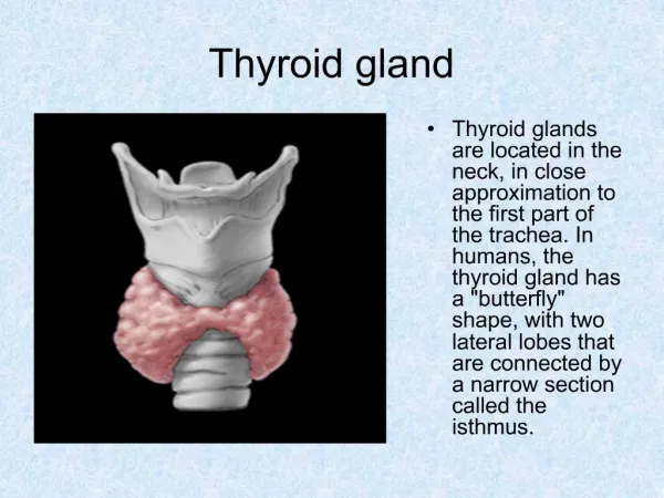
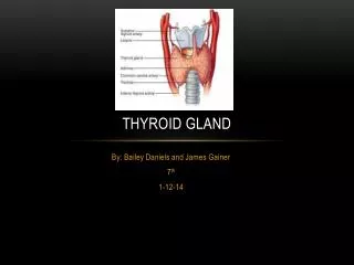
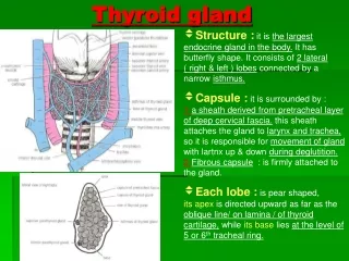
Thyroid gland pathology. By Dr.Mays Ibraheem. Pathology of thyroid gland including:. I. Hyperthyroidism. II. Hypothyroidism. III. Mass lesions of thyroid gland. Hyperthyroidism. Clinical features

E N D
Thyroid gland pathology By Dr.MaysIbraheem
Pathology of thyroid gland including: • I. Hyperthyroidism. • II. Hypothyroidism. • III. Mass lesions of thyroid gland.
Hyperthyroidism. Clinical features 1. Constitutional symptoms. Like soft, warm, flushed skin. Heat intolerance & excessive sweating are common. & weight loss despite increased appetite is due to sympathetic overactitivity. 2. Systematic symptoms. a. GIT symptoms. Include malabsorption syndrome, diarrhea. b. Cardiac symptoms. palpitation & tachycardia, congestive heart failure in elderly patients. c. Neuromuscular symptoms. Tremor, nervousness, & irritability, 50% develop muscle weakness. d. Ocular manifestations. Exopthalamus (wide staring gaze) and lid lag. e. Thyroid storm. It is a medical emergency characterized by abrupt onset of severe hyperthyroidism. This condition occurs most commonly in patient with Grave's disease. Significant number of patients dies because of cardiac arrhythmias.
The diagnosis based on clinical features & laboratory data. 1. Measurement of serum TSH concentration. The most useful single screening test for hyperthyroidism. • in primary hyperthyroidism (due to thyroid diseases), TSH levels are decreased even at the earliest stages • While in secondary hyperthyroidism (due to extra thyroid diseases like in diseases of pituitary or hypothalamus), TSH levels are either normal or high. 2. Measurement of free T4.
Grave's disease. (Autoimmune hyperthyroidism) • Most common cause of hyperthyroidism. • It is characterized by a triad of manifestations: • 1. Thyrotoxicosis • 2. Ophthalmopathy: exopthalamus in 40% of cases • 3. Pretibial myxedema • About 85% cases are occurred in young female between 20 – 40 years. • An increased incidence of Grave's disease occurs among family members (associated with HLA-DR3).
Pathogenesis of Grave's disease. Grave's disease is an autoimmune disease explanation:I. Presence of variety of autoantibodies in the serum of patient with grave's disease 1. Thyroid stimulating immunoglobulins (TSI) 2. Thyroid growth- stimulating immunoglobulins (TGI) 3. TSH binding inhibitor immunoglobulins (TBIIs) II. Presence many of CD4+ helper T cells within the thyroid. These cells are thought to trigger the B cells to produce autoantibodies.
Grave’s disease diffusely enlarged, soft, & its capsule is intact
There is diffuse hypertrophy & hyperplasia of thyroid follicular cells, which result in followings. 1. Follicular cells are crowded & become tall, columnar. 2. This crowding result in formation of papillae. 3. The colloid within the lumen of follicular lumen is pale, with scalloped margins. 4. Lymphoid infiltrates, consisting mainly of T cells, with fewer B cells & mature plasma cells; germinal centers are common.
Extrathyroidal changes. • Ophthalmopathy, the tissues of orbit are edematous, due to the presence of (hydrophilic glycosaminoglycans, infiltration by lymphocytes mostly T cells, & fibrosis of orbital muscles). • Dermatopathy is characterized by thickening of the dermis (deposition of glycosaminoglycans & lymphocyte infiltration).
Hashimoto thyroiditis • The most common cause of hypothyroidism where enough iodine is present Pathogenesis of Hashimoto thyroiditis. • I. CD8+ cytotoxic T cell–mediated cell death: CD8+ cytotoxic T cells may cause thyrocyte destruction(parenchymal destruction). • II. Binding of anti-thyroid antibodies followed by antibody-dependent cell-mediated cytotoxicity(. These antibodies are: • Anti-thyroid peroxidase antibody (anti-TPO). • Antithyroglobulinantibody (TgAb) • Inhibitory anti- TSH receptor antibodies • III. There is a significant genetic component to disease pathogenesis. By the following facts: • Increased frequency of disease in first degree relatives of patient. • An association has also been found between disease prevalence & the HLA subtypes DR3 & DR5.
Hashimoto’s disease Diffusely & symmetrical enlarged thyroid, the capsule of gland is intact & the gland is well demarcated from adjacent structure.
Microscopic features of Hashimoto’s disease Widespread infiltration of thyroid parenchyma by a mononuclear inflammatory infiltrate containing small lymphocytes, plasma cells, & well developed germinal centers. The thyroid follicles are atrophic & are lined by epithelial cells characterized by presence of abundant eosinophilic, granular cytoplasm, termed Hurthle (metaplastic cells) or Oxyphil cells.
Clinical features • More common in female than male, most prevalent between 45- 65 years of age. • Presents as painless enlargement of the thyroid gland, usually associated with some degree of hypothyroidism. • Patient with Hashimoto thyroiditis is at increased risk for the development of B- cells non-Hodgkin's lymphomas within the thyroid gland.