Download
1 / 16
450 likes | 1.26k Views
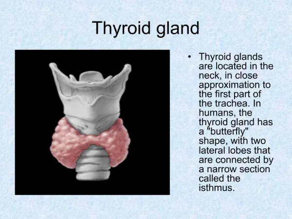
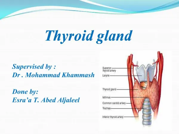
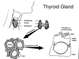
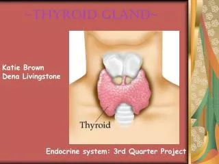
Thyroid Gland. Two types of cells are present in the follicle : Principal(follicular) cells Parafollicullar cells (C). The thyroid is an extremely vascularized organ Endothelial cells of these capillaries are fenestrated. . Thyroid Gland

E N D
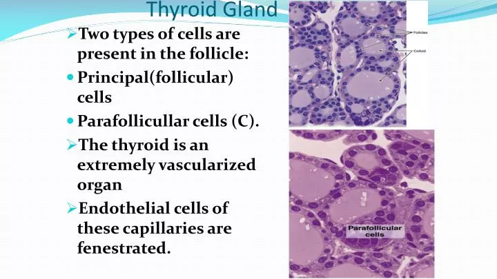
Thyroid Gland • Two types of cells are present in the follicle: • Principal(follicular) cells • Parafollicullar cells (C). • The thyroid is an extremely vascularized organ • Endothelial cells of these capillaries are fenestrated.
Thyroid Gland • The morphological appearance of thyroid follicles varies according to the region of the gland and its functional activity. • In the same gland: • Larger follicles that are full of colloid have a cuboidal or squamous epithelium . • Follicles with less colloid are lined by columnar epithelium.
Thyroid Gland • The parafollicular, or C, cell, is found as part of the follicular epithelium or as isolated clusters between thyroid follicles. • Parafollicular cells are larger than thyroid follicular cells and with the light microscope appear less stained they secrete calcitonin.
Parathyroid Glands • These are 4 glands. • Each parathyroid gland is contained within a connective tissue capsule. • These capsules send septa into the gland, where they merge with the reticular fibers that support elongated cordlike clusters of secretory cells • Cells of the Parathyroid • Chief cells. • Oxyphil.
Cells of theParathyroid: • Chief cells are small (pale,acidophilic). • Oxyphil are larger (darker,acidophilic). • Adipose tissue is found in large amounts with increase in age. • Chief cells secretes parathyroid hormone which promotes the absorption of the calcified bone matrix and the release of Ca2+ into the blood. • Oxyphil cells have PTH.
Endocrine Pancreas • The islets of Langerhans appear as rounded clusters of cells embedded within the exocrine pancreatic tissue they are more abundant in the tail of the pancreas. • A fine capsule of reticular fibers surrounds each islet, separating it from the adjacent pancreatic tissue.
Each islet consists of lightly stained rounded cells, arranged in cords separated by a network of blood capillaries . • The vascularization, composed of many fenestrated capillaries, is more extensive than that of the exocrine tissue.
Four types of cells A, B, D, and F have been recognized in the islets: • ~70% B or beta-cells which secrete insulin. Insulin stimulates the synthesis of glycogen, protein and fatty acids. • ~20% A or alpha-cells which secrete glucagon. • ~5% D or delta-cells which secrete somatostatin, inhibits other endocrine cells. • ~5% F which secrete pancreatic polypeptide
Insulin-dependent or type I diabetes (juvenile diabetes) results from partial or total destruction of B cells due to an autoimmune disease. • Insulin-independent diabetes or type II diabetes occurs at a later stage in life and is frequently associated with obesity.
Pineal Gland • The pineal gland is also known as the epiphysis cerebri, or pineal body. It is situated on the roof of the diencephalons. • The pineal gland is covered by pia mater.
Connective tissue septa containing blood vessels and unmyelinated nerve fibers penetrate the pineal tissue. • It also surrounds the cellular cords and follicles, forming irregular lobules. • The cells of pineal gland consists principally of: • Pinealocytes • Astrocytes. • The pinealocytes appear to have long and tortuous branches. • Pinealocytes produce melatonin and serotonin. • Brain sands (deposition of Mg and Ca)or corpora arenacea which characterize this gland are also present.
The astrocytes of the pineal gland are a specific type of cell characterized by elongated nuclei that stain more heavily than do those of parenchymal cells. • They are observed between the cords of pinealocytes and in perivascular areas. These cells have long cytoplasmic processes.