Download
1 / 64
640 likes | 651 Views
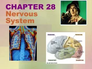
This chapter explains the stimulus-response process in the nervous system, discussing the structure and function of neurons, action potentials, synaptic transmission, and the integration of signals. It also covers common neurotransmitters and the condition multiple sclerosis.

E N D
stimulus Line of Communication receptors sensory neurons integrators interneurons motor neurons effectors muscles, glands response Fig. 34-2, p.574
Vertebrate Nervous Systems • Earliest fishlike vertebrates had a hollow, tubular nerve cord • Modification and expansion of nerve cord produced spinal cord and brain • Nerve cord persists in vertebrate embryos as a neural tube • Cephalization-formation of head and brain
Communication Lines Stimulus (input) Receptors (sensory neurons) Integrators (interneurons) motor neurons Effectors (muscles, glands) Response (output) Figure 34.5Page 575
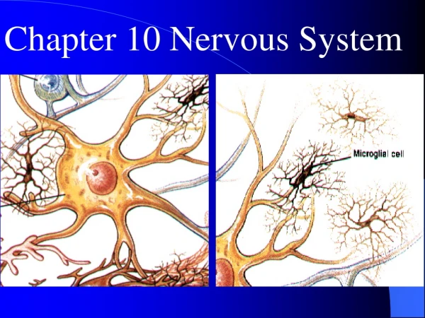
Neurons • Basic units of communication in nearly all nervous systems • Monitor information in and around the body and issue commands for responsive actions
Neurons Fig. 34-6d2, p.576
Motor Neuron dendrites Input Zone cell body axon axon endings Trigger Zone Conducting Zone Output Zone Stepped Art Fig. 34-6d1, p.576
Three Classes of Neurons • Sensory neurons • Interneurons • Motor neurons
dendrites axon cell body Fig. 34-6a, p.576
dendrites dendrites cell body axon Fig. 34-6b,c, p.576
Structure of a Neuron dendrites input zone cell body trigger zone conducting zone axon endings axon output zone Fig. 34-6d1, p.576
Resting Potential • Charge difference across the plasma membrane of a neuron • Fluid just outside cell is more negatively charged than fluid inside • Potential is measured in millivolts • Resting potential is usually about -70mv
How Ions Move across Membrane Interstitial fluid Cytoplasm Na+/K+ pump Passive transporters with open channels Passive transporters with voltage-sensitive gated channels Active transporters Lipid bilayer of neuron membrane Figure 34.7Page 577
Ion Concentrations at Resting Potential • Potassium (K+) • Higher inside than outside • Sodium (Na+) • Higher outside than inside
Na+ K+ outside plasma membrane inside Na+ K+ p.577
Action Potential • A transitory reversal in membrane potential • Voltage change causes voltage-gated channels in the membrane to open • Inside of neuron briefly becomes more positive than outside
Action Potential 1 Na+ 2 Na+ Na+ K+ K+ K+ K+ K+ K+ K+ Na+ Na+ Na+ Na+ 3 4 Na+ Na+ Figure 34.8a-dPage 578-79
Positive Feedback more Na+ ions flow into the neuron more gated channels for Na+ open neuron becomes more positive inside
All or Nothing • All action potentials are the same size • If stimulation is below threshold level, no action potential occurs • If it is above threshold level, cell is always depolarized to the same level
Repolarization • Once peak depolarization is reached, Na+ gates close and K+ gates open • Movement of K+ out of cell repolarizes the cell • The inside of the cell once again becomes more negative than the outside
electrode outside electrode inside unstimulated axon Fig. 34-9b, p.579
stimulated axon Fig. 34-9e1, p.579
action potential threshold level resting level Fig. 34-9e2, p.579
Propagation of Action Potentials • An action potential in one part of an axon brings a neighboring region to threshold • Action potential occurs in one patch of membrane after another
Chemical Synapse • Gap between the terminal ending of an axon and the input zone of another cell plasma membrane of axon ending of presynapic cell plasma membrane of postsynapic cell synaptic vesicle synaptic cleft membrane receptor Figure 34.10aPage 580
Synaptic Transmission • Action potential in axon ending of presynaptic cell causes voltage-gated calcium channels to open • Flow of calcium into presynaptic cell causes release of neurotransmitter into synaptic cleft
Synaptic Transmission • Neurotransmitter diffuses across cleft and binds to receptors on membrane of postsynaptic cell • Binding of neurotransmitter to receptors opens ion channels in the membrane of postsynaptic cell
Ion Gates Open neurotransmitter ions receptor for neurotransmitter gated channel protein Figure 34.10cPage 580
Synaptic Integration what action potential spiking would look like threshold -65 Membrane potential (milliseconds) EPSP integrated potential resting membrane potential -70 IPSP Figure 34.12Page 581 -75
neuromuscular junction motor neuron axons from spinal cord to skeletal muscle cells transverse slice of spinal cord part of a skeletal muscle Fig. 34-11a, p.581
axon ending muscle fiber Fig. 34-11b, p.581
Neurotransmitters • ACh • Norepinephrine • Epinephrine • Dopamine • Serotonin • GABA • Derived from amino acids
Multiple Sclerosis • A condition in which nerve fibers lose their myelin • Slows conduction • Symptoms include visual problems, numbness, muscle weakness, and fatigue
axon myelin sheath nerve fascicle Nerve • A bundle of axons enclosed within a connective tissue sheath Figure 34.15Page 584
Myelin Sheath • A series of Schwann cells • Sheath blocks ion movements • Action potential must “jump” from node to node Figure 34.15bPage 584
Reflexes • Automatic movements made in response to stimuli • In the simplest reflex arcs, sensory neurons synapse directly on motor neurons • Most reflexes involve an interneuron
Stretch Reflex STIMULUS Biceps stretches. sensoryneuron motorneuron Response Biceps contracts. Figure 34.16Page 585
Central and Peripheral Nervous Systems • Central nervous system (CNS) • Brain • Spinal cord • Peripheral nervous system • Nerves that thread through the body
Peripheral Nervous System • Somatic nerves • Motor functions • (Shown in green) • Autonomic nerves • Visceral functions • (Shown in red)
Function of the Spinal Cord • Expressway for signals between brain and peripheral nerves • Sensory and motor neurons make direct reflex connections in the spinal cord • Spinal reflexes do not involve the brain
Brain Development midbrain hindbrain forebrain Brain at 7 weeks Fig. 34-19b, p.588
Brain Development Brain at 9 weeks Fig. 34-19c, p.588
Brain Development Brain at birth Fig. 34-19d, p.588
right ventricle left ventricle third ventricle fourth ventricle spinal canal Fig. 34-20, p.588
Vertebrate Brains olfactory lobe olfactory lobe (part of forebrain) forebrain forebrain midbrain hindbrain midbrain hindbrain fish (shark) reptile (alligator) mammal (horse) Figure 34.21Page 589