Download
1 / 68
720 likes | 924 Views
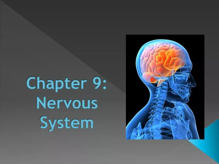
Chapter 9: Nervous System . Functions. Sensors Monitor external and internal environment Processing Receives information, integrates it, and decided what to do Effectors Carries messages to effectors and tells them what to do. Types of Nervous Cells. Neurons Main cell of nervous tissue

E N D
Functions • Sensors • Monitor external and internal environment • Processing • Receives information, integrates it, and decided what to do • Effectors • Carries messages to effectors and tells them what to do
Types of Nervous Cells • Neurons • Main cell of nervous tissue • Relay and process messages • Neuroglial • Provide support to the neurons • Several types known • Ex: Schwann, microglial
Neuroglial Cells • Microglial cells • Scattered throughout CNS • Phagocytize bacteria or cellular debris • Oligodendrocytes • Along nerve fibers • Provide myelin sheath (made of a fatty substance called myelin) around axon in CNS • Schwann cells • Same as oligodendrocytes but in PNS
Neuroglial Cells (cont) • Astrocytes • Provide connection between a neuron and a blood vessel • Provide support, help regulate ion concentrations in tissue, make-up scar tissue after injury • Ependymal cells • Forms epithelial-like linings on the outsides of specialized parts or lining cavities within the CNS
Neurons • Remember: It’s a cell! • Body of neuron • Cell Body – contains cell organelles • Dendrites- carry messages to cell body • Axons – carry messages away from cell body • Cell Structures • Large nucleus with easily seen nucleolus • Chromatophilic substance – similar to rough ER • Scattered throughout cytoplasm, membranous • Neurofibrils- help support cell shape
Axons • Can be myelinated or unmyelinated • PNS • Schwann cells form myelin sheath • Nodes of Ranvier- small breaks in myelin sheath • CNS • Oligodendrocytes form myelin • Myelinated neurons form white matter • Unmyelinated neurons form gray matter
Structural Classification of Neurons • Multipolar • Many small branched dendrites • One axon • Found in CNS • Bipolar • Two processes off of cell membrane (one axon and one dendrite) • Neurons in special sense organs • Unipolar • One process off of cell body (one axon) • Found throughout PNS
Functional Classification of Neurons • Sensory (afferent) neurons • Have sensitive dendrites that are stimulated by changes in environment • Message is taken into CNS • Usually unipolar or sometimes bipolar • Interneurons • Transfer, direct, and process messages within CNS • Usually multipolar • Motor (efferent) neurons • Carries message out of CNS to effectors • Usually multipolar
A Neuron at Rest • Inside the neuron • High in K+ • High in negative ions • Outside the neuron • High in Na+ • High in positive ions • Result • K+ tends to diffuse out • Na+ tends to diffuse in • Negative ions cannot cross
A Neuron at Rest (cont) • Na/K pump - helps to restore concentration gradient across the cell membrane • Resting potential - difference is electrical charge across the membrane • Established by concentration gradients of various ions • Inside of the membrane has a negative charge of 70 mv • Membrane is said to be polarized
Potential Changes • Stimuli cause changes to the resting potential by making the inside of the membrane less negative • Once a stimulus happens: • If stimulus is not strong enough to reach threshold potential = cell membrane will return to resting potential • If stimulus is strong enough to reach threshold potential = start an action potential • Summation - when additive effect of stimuli causes action potential
Action Potential • Starts at trigger zone of axon • Threshold stimulus open sodium channels • Sodium moves into axon • Because of the concentration gradient • Because of the negative charge that attracts the positive ions • Depolarizes the membrane as negative charge diminishes • Potassium channels open and potassium moves out of the axon, repolarizing the membrane • Animation #1Animation #2
Nerve Impulse • Action potential at the trigger zone stimulates the next part of the axon to do a action potential • Potentials spread along the axon like a wave • Unmyelinated axons • Wave continues uninterrupted; relatively slow • Myelinated axons • Wave goes through saltatory conduction (jump from one node to the next); very fast Animation
Neuron Responses • All-or-nothing effect • Neuron does not react until a threshold stimulus is applied, but once it is applied it reacts fully • Stimuli greater than threshold levels don’t change the size of the response but changes its frequency • Refractory period: • After a action potential • Brief period of time • The nerve cannot be stimulated again.
The Synapse • The connection between two neurons • Don’t touch, separated by synaptic cleft • One-way communication between axon of presynaptic neuron and dendrite of postsynaptic neuron • Neurotransmitters are made in the synaptic knob of the axon, stored in synaptic vesicles, and cross the cleft when needed
Neurotransmitter Actions • Excitatory Action: • A neurotransmitter that puts a neuron closer to an action potential (facilitation) or causes an action potential • Inhibitory Action: • A neurotransmitter that moves a neuron further away from an action potential • Response of neuron: • Responds according to the sum of all the neurotransmitters received at one time
Neurotransmitters • Acetylcholine • Monoamines – modified amino acids • Amino acids • Neuropeptides- short chains of amino acids • Depression: • Caused by the imbalances of neurotransmitters • Many drugs imitate neurotransmitters • Ex: Prozac, zoloft, alcohol, drugs, tobacco
Release of Neurotransmitters • When an action potential reaches the end of an axon, Ca+ channels in the neuron open • Causes Ca+ to rush in • Cause the synaptic vesicles to fuse with the cell membrane • Release the neurotransmitters into the synaptic cleft • After binding, neurotransmitters will either: • Be destroyed in the synaptic cleft OR • Taken back in to surrounding neurons (reuptake) • Animation
Neuronal Pools • Groups of highly interconnected neurons that work together in the CNS • Convergence • Axons from different parts of the nervous system connect to the same neuron combining their affects • Divergence • A message from one neuron is sent to many neurons at once; amplifies message
Divergence Convergence
Nerves • Nerves are made of bundled axons, called nerve fibers • Nerve fibers • Sensory (afferent)- carry messages to CNS • Motor (efferent)- carry messages from CNS to effectors • Nerves • Same definitions hold true • Most nerves contain both types of fibers and are called mixed nerves
Structure of a nerve • A nerve fiber (axon) is surrounded with endoneurium • Nerve fibers are bundled together and surrounded with perineurium to form a fascicle • Fascicles are bundled together and surrounded with epineurium to form a nerve
Nerve Pathways • Path that the message takes through the body • Includes: • Sensor • Sensory neuron • Interneuron • Motor neuron • Effector
Reflex Arc • Simplest nerve pathway is a reflex • Reflexes without pain • Involve only sensory and motor neurons • Ex: knee-jerk reflex • Reflexes with pain • Involve interneurons in CNS • Ex: withdrawal reflex
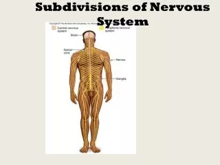
The CNS • Central nervous system • Consists of brain and spinal cord • Made of both gray and white matter • Covered in meningies
The Meninges (Brain) • Cranial Bone • Dura mater • First layer; tough, fibrous connective tissue • Forms inner periosteum of cranial bone • Folds into the cranium in some places to form division walls in the brain • Arachnoid mater • Web-like membrane over CNS • Does not dip into crevices
The Meninges (Brain) • Subarachnoid space • Below arachnoid layer • Contains cerebrospinal fluid • Pia mater • Lower layer of meninges • Forms a tight covering over brain • Does dip into crevaces
Meningies (Spinal Cord) • Same except: • Vertebrae bones - protection • Epidural space- filled with loose connective and adipose tissue • All other are the same
Cerebrospinal fluid • Flows through ventricles (spaces in brain) in the subarachnoid space, and through the central canal of the spinal cord • Fluid is made by the choroid plexus
Spinal Cord • Stretches from brain to intervertebral disk between first and second lumbar vertebrae • 31 pairs of spinal nerves come of the cord • Gray matter core surrounded by white matter
Spinal Cord (cont) • Responsible for communication between brain and body and spinal reflexes • Ascending tracts • Nerves that send info to brain • Descending tracts • Nerves that send into to effectors
Don’t forget: You can copy-paste this slide into other presentations, and move or resize the poll.
Don’t forget: You can copy-paste this slide into other presentations, and move or resize the poll.
The Brain • Made up off about 100 billion neurons • Four main sections: • 1) Cerebrum • Nerves for processing sensory and motor function • Higher functions (like reasoning) • 2) Diencephalon • Processes sensory information • 3) Brainstem • Regulates certain body functions like breathing • 4) Cerebellum • Coordinates skeletal muscle movements