Download
1 / 94
940 likes | 969 Views
Chapter 11: Nervous System. Function of the Nervous System To coordinate the actions of your body To ensure effective behaviour To maintain the internal environment within safe limits (homeostasis)

E N D
Function of the Nervous System • To coordinate the actions of your body • To ensure effective behaviour • To maintain the internal environment within safe limits (homeostasis) Messages are relayed throughout the body via electrochemical messages from the brain or through chemical messengers – hormones (hormones require more time than nervous transmission but are long lasting) There are more nerve cells in the body than there are visible stars in the Milky Way! 1 cm3 of brain tissue houses several million neurons with each connecting with several thousand others
Nervous Tissue The nervous system is divided into a central nervous system (CNS), consisting of the brain and spinal cord, and a peripheral nervous system (PNS), consisting of nerves carrying sensory and motor information between the CNS and muscles and glands. Both systems have two types of cells: neurons that transmit impulses and neuroglial cells that support neurons.
Neuron Structure Neurons are composed of dendrites that receive signals, a cell body with a nucleus, and an axon that conducts a nerve impulse away. Sensory neurons take information from sensory receptors to the CNS. Interneurons occur within the CNS and integrate input (nonmyelinated). Motor neurons take information from the CNS to muscles or glands.
dendrites – receive information (either from receptor cells or other nerve cells), conducting towards the cell body (~200 dendrites/cell body) cell body – location of the nucleus, high metabolic rate (so contains mitochondria) axon– may be 1m long, very thin, conducts the impulse towards other neurons or effectors, starts at axon hillock, the smaller the neuronal diameter, the faster the neuronal transmission
nodes of Ranvier– the unmyelinated sections of a myelinated neuron, impulses “jump” between the nodes of Ranvier neurilemma– a thin layer encompassing neurons in the peripheral nervous system, promoting their regeneration
Schwann cell – responsible for the myelin synthesis, type of glial cell (supporting and nourishing cell found in the nervous system) Axon Bulb – either at a synaptic bulb or end plate to muscle, contains neurotransmitter
Myelin Sheath Myelination covers long axons with a protective myelin sheath (made by neuroglial cells called Schwann cells). The sheath contains lipid myelin which gives nerve fibers their white, glistening appearance. The sheath is interrupted by gaps called nodes of Ranvier. Multiple sclerosis is a disease of the myelin sheath.
FYI Nerves are generally comprised of many neurons together (like fibre optic cable) Myelinated neurons in the brain are termed white matter (the myelin makes them look white) White matter may regenerate after injury, whereas grey matter (unprotected) will not
The Nerve Impulse The nervous system uses the nerve impulse to convey information. The nature of a nerve impulse has been studied by using excised axons and a voltmeter called an oscilloscope. Voltage (in millivolts, mV) measures the electrical potential difference between the inside and outside of the axon.
Membrane Polarization (Resting Potential) When an axon is not conducting a nerve impulse, the inside of an axon is negative (-70mV) compared to the outside(+40mV); this is the resting potential. To establish the –70mV potential in the cell: • Na+ is actively pumped out of the cell • K+ is actively pumped into the cell Sodium pump
Membrane Polarization (Resting Potential) • Na+ and K+ diffuse down the concentration gradient, but K+ diffuses faster due to an increased number of ion channels (gates) open to K+ ions • Since there is a net loss of positive ions to the outside of the cell, -70 mV is established inside the neuron • There are also large negative proteins inside the neuron that contribute to the negative charge
Membrane Depolarization When the nerve cell is excited, the membrane DEPOLARIZES (Action Potential) The membrane’s polarity changes: – Na+ channels open, Na+ rushes in, K+ gates close The positive ions flowing in causes a charge reversal to +40 mV inside the neuron (gated channel proteins)
Membrane Repolarization Once the charge becomes positive, the Na+ gates close, K+ gates open, eventually restoring the charge inside the neuron to –70 mV (but the Na+ excess is inside and K+ excess is outside!) The Na/K Pumprestores the ion concentrations inside and outside the cell
Membrane Repolarization During the repolarization, the nerve cannot be reactivated – this is called the refractory period (1 to 10 ms) and is a recovery time for the neuron The pump requires ATP in order to operate
The Na/K Pump To be ready for another action potential, the membrane re-establishes the proper concentration gradient for sodium and potassium Three sodium ions are actively transported across the membrane and to the ECM Two potassium ions are then carried across to the cytoplasm
Movement of the Action Potential The action in the neuron adjacent to an area of resting membrane causes that area to depolarize, moving the action potential along (due to attraction of opposite charges) Since the area from which the action potential came is still in recovery, the action potential will only move in one direction
Propagation of an Action Potential The action potential travels the length of an axon, with each portion of the axon undergoing depolarization then repolarization. A refractory periodensures that the action potential will not move backwards. In myelinated fibers, the action potential only occurs at the nodes of Ranvier. This “jumping” from node-to-node is called saltatory conduction.
Fig. 48-13 Schwann cell Depolarized region (node of Ranvier) Cell body Myelin sheath Axon
The All-or-None Response (Threshold Potential) All neurons provide an all-or-none response: - in response to a stimulus, they either activate (fire) and provide a certain level of response, or don’t fire at all A neuron will only fire if it is stimulated with an intensity of at least threshold level Every action potential for a neuron is identical in strength and duration (regardless of how much beyond threshold the stimulus is)
Threshold Potential All neurons differ in their threshold level To inform the brain of the intensity of a stimulus: - the frequency of firing is increased (not speed, which is constant for each neuron) - the number of neurons that respond to that level of stimulus can increase (neurons may have different threshold)
Transmission Across a Synapse • The junction between neurons or neurons & effectors is called the synapse. • Transmission of a nerve impulse takes place when a neurotransmitter molecule stored in synaptic vesicles in the axon bulb is released into a synaptic cleft between the axon and the receiving neuron.
When a nerve impulse reaches an axon bulb, calcium channels open and Ca2+ flow into the bulb. This sudden rise in Ca2+ causes synaptic vesicles to move and merge with the presynaptic membrane, releasing their neurotransmitter molecules into the synapse The binding of the neurotransmitter to receptors in the postsynaptic membrane causes either excitation or inhibition.
Synaptic Summation Many synapses per single neuron is not uncommon. Excitatory signals have a depolarizing effect, and inhibitory signals have a hyperpolarizing effect on the post- synaptic membrane. Summation is the summing up of these excitatory and inhibitory signals.
Neurotransmitter Molecules Out of 25, two well-known neurotransmitters are acetylcholine (ACh) and norepinephrine (NE). Neurotransmitters that have done their job are removed from the cleft; the enzyme acetylcholinesterase(AChE) breaks down acetylcholine. Neurotransmitter molecules are removed from the cleft by enzymatic breakdown or by reabsorption, thus preventing continuous stimulation or inhibition.
FYI • most synapses involve more than just 2 neurons (or neuron/effectors) • neurotransmitters move only by diffusion, so synaptic transmission is MUCH slower than axonal transmission. • insecticides interfere with enzymes that break down neurotransmitters causing their hearts to remain contracted, • whereas LSD and other hallucinogens are believed to bind to the receptor sites for neurotransmitters
Lidocaine, an anesthetic works by stabilizing the neuronal membrane so it can’t depolarize • Endorphins and enkephalins are “natural” painkillers produced in the CNS, blocking the pain transmitter that usually attaches to the injured organ allowing the perception of pain • opiates (heroin, codeine, morphine) block the production of the pain transmitter. Since they act to decrease the production of natural painkillers, the amount of opiate taken must be increased or at least maintained to maintain the effect
Valium and other depressants are believed to enhance the action of inhibitory synapses • Alcohol acts to increase the polarization of the membrane, increasing the threshold • Since many neurons will connect to a postsynaptic neuron, it is the summation of the effects of the presynaptic neurons that determine whether or not the postsynaptic neuron or effector will depolarize
Neural Circuits – includes neuronal and synaptic transmission There are two types of neural circuits • complicated neural circuits, involving conscious thought • reflex arcs – without brain coordination • often unconscious, involuntary and faster than when thought is required (why are these useful?)
Nervous Control (in general) StimulusReceptorSensoryNeuronInterneuron BrainInterneuronMotorNeuronEffectorResponse Reflex Arc (see diagram – the reflex arc) StimulusReceptorSensoryNeuronInterneuron (spinal cord)MotorNeuronEffectorResponse When the response is made at the spinal cord level (information does not have to go to the brain to be processed), the response is quick (and always correct given the circumstances) Reflexes protect the body from injury
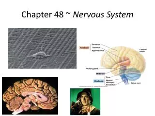
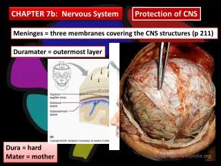
The Central Nervous System The central nervous system (CNS) consists of the spinal cord and brain. Both are protected by bone, wrapped in protective membranes called meninges, and surrounded and cushioned with cerebrospinal fluid that is produced in the ventricles of the brain.
The ventricles are interconnecting cavities that produce and serve as a reservoir for cerebrospinal fluid. The CNS receives and integrates sensory input and formulates motor output. Gray matter contains cell bodies and short, nonmyelinated fibers; white matter contains myelinated axons that run in tracts.
The Brain • consumes more oxygen and glucose than any other part of the body • meninges – outer layers (protection) – dura mater, arachnoid and pia mater • cerebrospinal fluid –between the inner, middle meninges & central canal of s.cord, carries nutrients, acts as a shock absorber, relays waste by diffusion & fac. diffusion, flows within ventricles – four “spaces” in the brain
Fig. 49-15 Frontal lobe Parietal lobe Somatosensory cortex Motor cortex Somatosensory association area Speech Frontal association area Taste Reading Speech Hearing Visual association area Smell Auditory association area Vision Temporal lobe Occipital lobe
Fig. 49-17 Max Seeing words Hearing words Min Speaking words Generating words
The Cerebral Cortex The cerebral cortex is a thin, highly convoluted outer layer of gray matter covering both hemispheres. The primary motor area is in the frontal lobe; this commands skeletal muscle. The primary somatosensory area is dorsal to the central sulcus or groove.
Forebrain (cerebrum) • contains two hemispheres for coordinating sensory and motor information – speech, reasoning, memory, personality, which may be located on one side only • the outer layer is called the cerebral cortex (only 1 mm thick), deeply folded into fissures(to increase surface area)