Download
1 / 90
900 likes | 903 Views
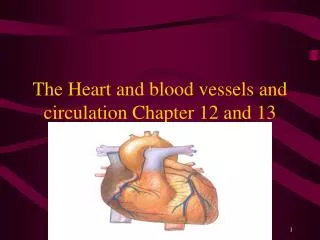
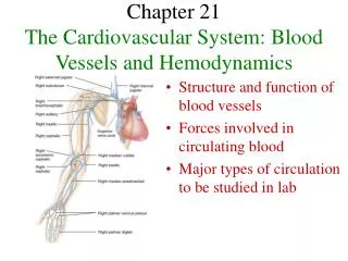
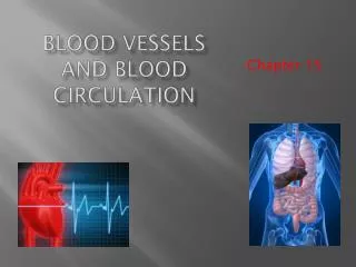
Chapter 21 Blood Vessels and Circulation. BIO 211 Lab Instructor Dr. Gollwitzer. Today in class we will discuss: General characteristics of blood vessels The anatomy of blood vessels The differences between arteries and veins The general functional patterns of blood vessels

E N D
Chapter 21 Blood Vessels and Circulation BIO 211 Lab Instructor Dr. Gollwitzer
Today in class we will discuss: • General characteristics of blood vessels • The anatomy of blood vessels • The differences between arteries and veins • The general functional patterns of blood vessels • The types of arteries based on size, structure, and basic function • Cardiovascular receptors • And identify the major systemic arteries of the: • Aortic arch • Head, brain, and neck • Upper and lower extremities • Trunk (thoracic and abdominal regions)
Blood Vessels • Arteries (a=away from heart) arterioles capillaries venules veins (to heart) • Arteries branch repeatedly, decrease in diameter • Arterioles smallest branches of arteries; deliver blood to capillaries • Capillaries smallest, thinnest vessels • Site of all chemical and gaseous exchange between blood and interstitial fluid occurs • Tissues rely on capillary diffusion to obtain nutrients and oxygen and to remove carbon dioxide and metabolic wastes • Venules smallest venous branches; collect blood from capillaries; unite to form veins • Veins return blood to heart • Arteries and veins typically lie side by side in region being served
Anatomy of Blood Vessels • 3 layers to vessel walls • Tunica interna/intima innermost layer • Endothelium and CT with elastic fibers • In arteries outer margin contains internal elastic membrane (thick layer of elastic fibers) • Tunica media middle layer • Concentric sheets of smooth muscle with loose CT • When smooth muscle contractsdiameter decreases; when relaxesincreases • In arteries • Thickest layer • Outer margin contains external elastic membrane (thin band of elastic fibers) • Larger arteries also contain layers of longitudinal smooth muscle cells • Tunica externa outermost layer • CT sheath around vessel • In arteries contains collagen and elastic fibers • In veins generally thicker than tunica media, contains elastic fibers and smooth muscle cells
Differences Between Arteries and Veins • Arteries more resilient • When stretched, elongate, keep shape; snap back when released • Veins contain valves to prevent backflow of blood toward capillaries
Vasa Vasorum • “Vessels of vessels” • Walls of large vessels • Too thick to allow diffusion between blood and blood vessel tissue • Contain small arteries and veins that supply smooth muscle cells and fibroblasts of tunica media and tunica externa
General Functional Patterns • Distribution of arteries and veins on L and R sides usually identical • L and R subclavian, axillary, brachial and radial arteries parallel veins • NOT parallel near heart where largest vessels connect to atria or ventricles • Single vessel may have several names as it crosses anatomical boundaries • Descending aorta, thoracic aorta, abdominal aorta • External iliac artery becomes femoral artery as it leaves the trunk and enters the lower limb
Structural Characteristics of Vessels Change Gradually as They Travel Away from/Toward Heart Figure 21-2
Arteries • Thick, muscular walls, make arteries elastic and contractile • Elasticity • Permits passive changes in vessel diameter in response to pressure waves that come with each heart beat • Allows arteries to absorb pressure pulses that accompanies contractions of ventricles • Contractility ability to reduce diameter; actively under control of sympathetic ANS • Vasoconstriction = contraction of arterial smooth muscle; decreases vessel diameter • Vasodilation = relaxation of arterial smooth muscle; increases vessel diameter • Important in hemostasis; contract to stop bleeding
Arteries • From heart to capillaries, arteries change • From elastic arteries (large arteries) • To muscular arteries (medium-sized arteries) • To arterioles
Elastic Arteries • AKA: conducting arteries • Large vessels • Transport large volumes of blood away from heart (e.g., pulmonary trunk, aorta, and major branches) • Walls very resilient • Tunica media contains many elastic fibers and few smooth muscle cells • Tolerate pressure changes during cardiac cycle • Increase BP arteries expand • Decrease BP arteries recoil to original shape • Elasticity dampens pressure peaks and valleys that accompany the heartbeat
Muscular Arteries • AKA: distribution arteries • Medium-sized vessels; are majority of arteries • Distribute blood to skeletal muscle and internal organs (e.g., external carotid arteries of neck, brachial arteries of arms, femoral arteries of legs) • Thick tunica media with more smooth muscle than elastic fibers • By the time blood reaches arterioles • Pressure oscillations have disappeared • Blood flow is continuous
Arterioles • Smallest arterial vessels • Have little or no tunica externa • Tunica media • In larger arterioles, only one or two layers of smooth muscle cells • Smallest arterioles have scattered smooth muscle cells • Respond to local conditions or to sympathetic or endocrine stimulation • Vasodilate when O2 levels are low (passive) • Vasoconstrict under sympathetic control • Changes in diameter affect amount of force needed to push blood around CV system • More pressure required to push blood though a constricted vessel than through a dilated one • RESISTANCE = force opposing blood flow • Arterioles called resistance vessels
Cardiovascular Receptors • 2 Types: • Baroreceptors • Chemoreceptors • Chemoreceptors • Respond to changes in O2, CO2, pH in blood • Located in • Carotid bodies (near carotid sinuses) • Aortic bodies (near aortic arch)
Cardiovascular Receptors • Baroreceptors • Located in • Carotid sinuses (expanded chambers near base of internal carotid arteries) (Fig 21-23) • Aortic sinuses (sac-like dilations at base of ascending aorta) (Fig 20-8b) • Monitor amount of stretch in walls of • Arteries • R atrium • Reflexes adjust CO and peripheral resistance to maintain normal arterial pressures • Inc BP dec CO and peripheral vasodilation
Locations of Cardiovascular Receptors Figure 20-8b Figure 21-23
Aneurysm • = Bulge in weakened wall of artery • Just like a bad tire can suffer a blow out • Most dangerous in • Arteries in brain stroke • Aorta fatal bleeding • Often painless, so go undetected • Occurrence • in individuals with arteriosclerosis (= thickening or toughening of vessel walls); over time vessels become less elastic and weak spot develops • in individuals with high BP; puts great stress on vessel walls • arterial inflammation or infection weakens arterial walls
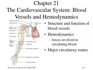
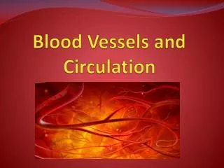
Major Systemic Arteries Figure 21-21
Systemic Arteries • Ascending aorta • Begins at aortic valve of left ventricle • R and L coronary arteries (branch from base) • Curves to form aortic arch • Turns downward to become descending aorta • Thoracic aorta • Abdominal aorta • Aortic arch • L subclavian artery • Vertebral • Axillary • L common carotid artery • Brachiocephalic trunk/artery • R common carotid artery • R subclavian artery
Arteries of the Head and Neck • Common carotid arteries ( head, neck) • Each divides into • External carotid artery • neck, lower jaw, esophagus, larynx, pharynx, face • Internal carotid artery • brain, cranial nerves • cerebral arterial circle/of Willis = ring of arteries that encircles infundibulum of pituitary gland • Circle is present because internal carotid arteries, middle cerebral arteries, and basilar artery are interconnected by anterior and posterior cerebral arteries and the anterior and posterior communicating arteries
Cardiovascular Receptors • Baroreceptors • Monitor degree of stretch in walls of arteries and right atrium • Located in carotid sinuses (expanded chambers near base on internal carotid arteries) and aortic sinuses (pockets in walls of ascending aorta adjacent to heart and wall of right atrium near entrance to vena cavae) • Reflexes adjust CO and peripheral resistance to maintain normal arterial pressures • Inc BP dec CO and peripheral vasodilation Figure 21-23
Arteries of the Brain • Originate from • Internal carotids (from common carotids) • Vertebrals (from subclavians) • Connected blood supply to brain • Internal carotid arteries normally supply anterior half of cerebrum • Vertebral arteries usually supply rest of brain • Are interconnected (via anastomoses) ring of arteries around base of brain = cerebral arterial circle (Circle of Willis) • Encircles hypothalamus, influndibulum, pituitary • Brain can receive blood from several vessels, so possibility of interruption of circulation is reduced (= collateral circulation)
Cerebral Arterial Circle(Circle of Willis) Figure 21-24a
Strokes • aka cerebrovascular accidents (CVAs) • Usually affect middle cerebral arteries • On right side loss of sensation and motor control on left side of body; difficulty drawing or interpreting spatial relationships • On left side aphasia and sensory and motor paralysis of right side of body • Strokes affecting vessels that supply the brain stem also have distinctive symptoms • Those affecting the lower brain stem are often fatal
Arteries of the Chest andUpper Limbs • Subclavian arteries • Branch in thoracic cavity (Fig 21-24a) • Vertebral arteries ( brain and spinal cord) • Thyrocervical trunks ( muscles of neck, shoulder, upper back) • Internal thoracic arteries ( pericardium and anterior chest wall) • Leave thoracic cavity • Axillary arteries (at axilla) • Brachial arteries (as they enter brachium) • Radial and ulnar arteries • Radial and ulnar merge at wrist • Palmar arterial arches • digital arteries capillaries
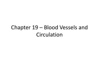
Arteries of the Trunk Figure 21-25
Arteries of the Trunk • Descending aorta • Divided by diaphragm into • Thoracic aorta • Abdominal aorta
Thoracic Aorta • Begins at T5 and ends at diaphragm (T12) • Visceral branches supply organs of chest • Bronchial arteries tissues of lungs NOT involved in gas exchange • Pericardial arteries pericardium • Esophageal arteries esophagus • Mediastinal arteries tissues of mediastinum • Parietal branches supply chest wall • Intercostal arteries chest muscles, vertebral column area • Superior phrenic arteries superior surface of diaphragm
Abdominal Aorta • Abdominal aorta (from inferior diaphragm to L4) • Unpaired branches • to visceral organs • Paired branches • to body wall • kidneys • urinary bladder
Arteries of the Abdominopelvic Organs Figure 21-25
Arteries of the Trunk Figure 21-25
Unpaired Branches of the Abdominal Aorta • Celiac trunk, divides into • left gastric artery stomach, inferior esophagus • splenic artery spleen, stomach, pancreas • common hepatic artery liver (through proper hepatic artery, then the hepatic arteries), stomach, gall bladder, duodenum, pancreas • Superior mesenteric artery pancreas, small intestine, appendix, upper 2/3 large intestine • Inferior mesenteric artery last 1/3 large intestine
Paired Branches of the Abdominal Aorta • Inferior phrenic arteries inferior surface of diaphragm, inferior esophagus • Suprarenal arteries adrenal glands • Renal arteries kidneys • Gonadal arteries gonads • In males = testicular arteries • In females = ovarian arteries • Lumbar arteries vertebrae, spinal cord, abdominal wall
Arteries of the Trunk Figure 21-25
Distal Branches of theAbdominal Aorta • Abdominal aorta divides at terminal segment (L4) into • L & R common iliac artery • Common iliac artery • Internal iliac artery pelvic structures and organs • External iliac artery lower limb
Arteries of the Lower Limb • External iliac arteries • Pass through abdominal wall • In thigh • Femoral arteries • deep femoral artery (skin and deep muscles of thigh) • Posterior to knee • Popliteal artery • Anterior tibial artery dorsalis pedis ( foot/ankle) • Posterior tibial artery • Fibular (peroneal) artery (at ankle) • Plantar arteries (plantar surface of foot) • Plantar arteries and dorsalis pedis arteries anastomose to form plantar arterial arches • Digital artery arterioles capillaries
Today in class we will discuss: • The structure of capillaries, different types of capillaries, and they function • The types of veins based on size, structure, and basic function • Blood distribution among the different type of blood vessels • And identify the major systemic veins associated with: • Head, brain, and neck • Upper and lower extremities • Trunk (thoracic and abdominal regions) • The hepatic portal system
Capillaries • Microscopic capillary networks in ALL active tissues; no cell more than 5 cells from a capillary • Thin walls permit 2 way exchange/diffusion between blood and interstitial fluids; short distances, quick exchange • Slow blood flow sufficient time for diffusion or active transport of materials across walls