Download
1 / 63
700 likes | 1.01k Views
Chapter 20– Blood Vessels & Circulation. Ch. 20 (Blood vessels) Study Guide. Critically read Chapter 20 pp. 756-774 right before 20.4 “Venous return and circulatory shock” section. Also read Table 20.3 (p.782) and Insight 20.5 (p.808) in the textbook. Comprehend Terminology (those in bold)

E N D
Chapter 20– Blood Vessels & Circulation
Ch. 20 (Blood vessels) Study Guide Critically read Chapter 20 pp. 756-774 right before 20.4 “Venous return and circulatory shock” section. Also read Table 20.3 (p.782) and Insight 20.5 (p.808) in the textbook. Comprehend Terminology (those in bold) Study-- Figure questions, Think About It questions, and Before You Go On (section-ending) questions Do Testing Your Recall— 1-8, 10-12, 14-16, 18 Do True or False– 1-2, 4-5, 8-9 Do Testing Your Comprehension-- #5 2
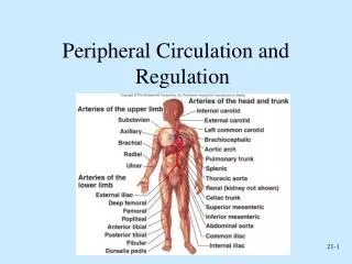
§ Introduction of Blood Vessels 1A. Closed circulatory system– Def. Blood flows in a continuous circuit through the body under pressure generated by the heart. • 1B. Open circulatory system-- In what animals? 2. Three principal categories of blood vessels: • Arteries: efferent vessels • Capillaries: • Veins: afferent vessels Fig. 20.x
§ Vessel wall of arteries/veins-1 1. Innermost layer (tunica interna/intima) A. Structures: lines the inside of the vessel and is exposed to the blood; consists of-- • Endothelial cells– histology? • Basement membrane • Connective tissue (sparse) B. Functions of the endothelial cells— • Selectively permeable barrier • Secrets chemicals--? • Repels blood cells and platelets Fig. x
The vessel wall 1 2 Next slide 3
§ Vessel wall of arteries/veins-2 2. Middle layer (tunica media)—thickest layer • Structures: • Smooth muscle cells-- • Collagen fibers • Elastic fibers (in arteries) B. Functions of this layer: • Strengthen the vessel • Provide vasomotion--?
§ Vessel wall of arteries/veins-3 3. Outermost layer (tunica externa or advertitia)— A. Structures: • Largely loose connective tissue (collagen fibers) B. Functions: • Protection & anchoring • Provide passage for-- • Vasa vasorum— vessels of the vessels Fig. 20.2
Conducting (large) artery Lumen Tunica interna: Endothelium Basement membrane Tunica media Tunica externa Vasa vasorum Nerve
§ Arteries • More muscular • Able to resist high blood pressure • Thus called resistance vessels • Retain their round shape even when empty • Divided into three categories by size (next slide)
§ Categories of Arteries-1 1. Conducting (elastic/large) arteries - largest • Ex. aorta, common carotid, subclavian, common iliac, and pulmonary trunk (Fig. 20.23) • Structure– (Slide #10) • tunica media-- 40-70 layers of smooth muscle alternating with elastic tissue • Internal/external elastic lamina— not obvious • tunica externa– vasa vasorum • Function-- • Able to expand/recoil-- • But not so in atherosclerosis– aneurysms and rupture (Slides #15-16)
L. common carotid a. R. common carotid a. R. subclavian a. L. subclavian a. Brachiocephalic trunk Aortic arch Ascending aorta Descending aorta, thoracic (posterior to heart) Diaphragm Aortic hiatus Descending aorta, abdominal 20-14
Aneurysm (read p. 758 box) • Def.– a balloon-like outpocketing of an artery wall (Fig. Y) • Risk– for rupture, most often reflects gradual weakening of the artery • Causes– OFTEN chronic hypertension or atherosclerosis • Common sites– abdominal aorta, renal arteries, and the arterial circle at base of brain Fig. Y
§ Categories of Arteries-2 2. Distributing (muscular, medium) arteries • Distribute blood to specific organs • Ex. brachial, femoral, renal, and splenic arteries etc. • Structure-- • tunica media– up to 40 layers of smooth muscle • Internal/external elastic lamina— conspicuous/not conspicuous (circle one) Fig. 20.34, 29, 30, 36
§ Categories of Arteries-3 3. Resistance (small) arteries • Up to 25 layers of smooth muscle • Elastic tissue little • ARTERIOLES (smallest of these); 1-3 smooth m. layers • Empty blood into capillaries through ____________________ • Here individual muscle cells form a precapillary sphincter encircling the entrance to capillary; function? Fig. 20.3
1 2 2a 3 4
§ Arterial Sense Organs (3 kinds) • Where– structures in major arteries above heart • Function– to monitor blood pressure/chemistry Three kinds (2 categories): Fig. 20.4 • Carotid sinuses (Baroreceptors)—Details next • Location-- in walls of ascending aorta etc. • monitors BP – a rise in BP signals brainstem . . . • Carotid bodies (Chemoreceptors) • Location-- oval bodies near carotids • monitor blood chemistry • adjust respiratory rate to stabilize pH, CO2, and O2 • Aortic bodies (Chemoreceptors) • Location-- in walls of aortic arch • same function as carotid bodies
Figure 20.4 2 To brain 1C To the face 3 1A+B
§ Capillaries • Material exchanges– between blood and tissue fluids • Locations-- _____________ and smallest of the venules • Structure– endothelium + ____________ • Fig. X next • Close vicinity to all cells— Exceptions • Scarce in: tendons, ligaments, & cartilage • Absent from (3 locations): -__________________________(Epi. & Eyes)
§ 3 Types of Capillaries 1. Continuous capillaries- occur in most tissues, ex. Skeletal muscle • endothelial cells have tight junctions with intercellular clefts (allow passage of solutes) • What molecules can pass– ex. glucose • What molecules can not– protein, formed elements of the blood • Fig. 20.5
§ 3 Types of Capillaries 2. Fenestrated capillaries • Structure – have _____________ on endothelial cells • filtration pores – spanned by very thin glycoprotein layer - allows passage of molecules such as _____________ • Locations-- organs that require rapid absorption or filtration - kidneys, small intestine etc. • Fig. 20.6 a and b
§ 3 Types of Capillaries 3. Sinusoids (discontinuous) capillaries- • Structure– endothelial cells separated by wide gaps; no basal lamina • Conform to the shape of the surrounding tissue • Molecules can pass– proteins and blood cells • Locations-- liver, bone marrow, spleen, lymphatic organs • Fig. 20.7
§ Veins (capacitance vessels; why?) • b/c Greater capacity for blood containment than arteries do (Fig. 20.8) • thinner walls—due to less muscular and elastic tissue; why? • lower blood pressure: 10 mm Hg with little fluctuation • ____________ aid skeletal muscles in upward blood flow
§ Types of veins-- Smallest to largest vessels (A) • Postcapillary venules-- only tunica intima • Receive blood from capillaries • more porous than capillaries • Muscular venules-- receive blood from #1 • have tunica media (1-2 layers of smooth muscle) + thin tunica externa • Medium veins– • Most have individual names, Examples-- radius or ulna veins • Many have venous valves
§ Types of veins-- Smallest to largest vessels (B) • Venous sinuses-- • veins with thin walls, large lumens, no smooth muscle; vasomotion– yes/no? (Circle one) • Examples– coronary sinus of the heart and the dural sinuses of the brain • Large veins-- • Greater than 10 mm (diameters) • Venaecavae, pulmonary veins, internal jugular veins
§ Circulatory Routes • Most common route • heart arteries arteriolescapillariesvenules veins • Portal system • blood flows through two consecutive capillary networks before returning to heart • 3 places in human body–
B. Hepatic portal sys. (p.797) Figure 20.38b 4 3 5 2 1
§ 3 Anastomoses Def. Point where 2 blood vessels merge • Arteriovenous shunt • artery flows directly into vein; fingers etc. • Venous anastomosis • most common type • alternate drainage of organs; Fig. 20.33 • Arterial anastomosis • Two arteries merge • collateral circulation (coronary); Fig. 20.31
What type of circulatory route does inferior/superior mesenteric vein belong?
§ Blood pressure, resistance, and flow • Importance– deliver oxygen and nutrients and to remove wastes at a rate keeps pace with tissue metabolism • Blood flow (F)– is the amount of blood flowing through an organ, tissue, or blood vessel in a given time • Hemodynamics:Blood Flow (F) = ΔP/R • Where ΔP is the pressure difference and R is the resistance
§ Blood Pressure • Blood pressure (BP)– Def.the force per unit area exerted by the blood against a vessel wall • In what vessels can you find BP? Figure 20.10 has the answer
Why ? Why?
§ Blood Pressure • BP is understood to mean the pressure in the _________________ • BP rises and falls in a pulsatile fashion in the arteries and arterioles Figure Z (what BP do we measure?)
Four different kinds of arterial BP-- A--? C. D. Mean arterial pressure B--?
§ Blood Pressure • Systolic P.–the maximum p. exerted in the arteries when blood is ejected into themduring ventricular ejection, averages 120 mm Hg (Mercury) • Physiology– during ventricular systole, a volume of blood enters the arteries from the ventricle. How much actually moves to the arterioles? • Status of the semilunar valves in this particular cardiac cycle? (open or close)
§ Blood Pressure • Diastolic P.– the arterial p. when blood is draining off into the arterioles during diastole, averages ________ Hg. Lowest during cardiac cycle. • Physiology–during ventricular diastole, the semilunar valves close, no blood enters the arteries but the arteries moves the blood forward. Why?
§ Blood Pressure • Pulse P.– is the difference between systolic and diastolic pressure • The Mean Arterial P. (MAP)— is the average blood pressure throughout the cardiac cycle • is monitored and regulated by BP reflexes • MAP = diastolic p. + 1/3 pulse p. Figure Z
Fig.-- Aortic pressure throughout the cardiac cycle Systolic pressure Mean arterial pressure Diastolic pressure A.
Q.—Peter’s systolic pressure is 140 mm Hg and his diastolic pressure is 95 mm Hg (written 140/95). A) What is his systolic p. and diastolic p., respectively? B) His pulse pressure? C) His mean arterial pressure?
§ Hypertension/hypotension Def.– high blood pressure; a chronic resting blood pressure higher than 140/90-- (hypertension) Results– aneurysms, atherosclerosis, heart failure, stroke, etc. Hypotension– a chronic low resting BP (90/50 or lower); Causes– blood loss, dehydration, anemia, in people approaching death