Download
1 / 40
430 likes | 1.28k Views
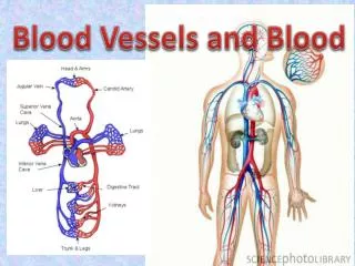
Chapter 14: Blood Vessels and Blood Circulation. The Vascular System. Closed system Blood vessels Four heart chambers. Blood Vessels. Five types of blood vessels Arteries Arterioles Capillaries Venules Veins. Blood Circuits. Two groups of blood vessels The pulmonary circuit

E N D
The Vascular System Closed system • Blood vessels • Four heart chambers
Blood Vessels Five types of blood vessels • Arteries • Arterioles • Capillaries • Venules • Veins
Blood Circuits Two groups of blood vessels • The pulmonary circuit • Pulmonary artery and its branches • Capillaries in lungs • Pulmonary veins • The systemic circuit • Aorta • Systemic capillaries • Systemic veins
Vessel Structure Three tunics (coats) of arteries and veins • Inner (endothelium) • Middle (smooth [voluntary] muscle) • Controlled by autonomic nervous system • Thinner in veins • Outer (supporting connective tissue)
Systemic Arteries The aorta • Largest artery • Receives blood from left ventricle • Branches to all organs
The Aorta and Its Parts • Ascending aorta • Aortic arch • Thoracic aorta • Abdominal aorta
Branches of the Ascending Aorta and Aortic Arch • Ascending aorta • *Left and right coronary arteries • Aortic arch • Brachiocephalic artery • Right subclavian artery • *Right common carotid artery • Left common carotid artery • Left subclavian artery
Branches of the Thoracic Aorta • *Branches to chest wall, esophagus, and bronchi • Intercostal arteries
Branches of the Abdominal Aorta • Celiac trunk • Left gastric artery • Splenic artery • Hepatic artery • Superior mesenteric artery • Small intestine • Inferior mesenteric artery • **Paired lateral branches • Phrenic arteries • Suprarenal arteries • Renal arteries • Ovarian and testicular arteries • Lumbar arteries
The Iliac Arteries and Their Subdivisions • Internal iliac arteries • two • External iliac arteries • Femoral artery • Popliteal artery • Tibial arteries • Dorsalis pedis
Arteries That Branch to the Arm and Head • External carotid artery • Internal carotid artery • Subclavian artery • Vertebral artery • Axillary artery • Brachial artery • Radial artery • Ulnar artery
Anastomoses Communication between two vessels • Circle of Willis • Superficial palmar arch • Mesenteric arches • Arterial arches
Arteries that supply the brain. The bracket at right groups the arteries that make up the circle of Willis.
Capillary network showing an arteriovenous shunt (anastomosis).
Systemic Veins • **Superficial veins • Cephalic, basilic, median cubital veins • Saphenous veins-longest in body • **Deep veins • Femoral and iliac vessels • Brachial, axillary, subclavian vessels • Jugular veins-two, drain carotids • Brachiocephalic vein-formed by the union of the subclavian and jugular veins
The Venae Cavae and Their Tributaries • Superior vena cava • Head, neck, upper extremities • Azygos vein • Chest wall • Inferior vena cava • Right, left veins from paired parts, organs such as the iliacs and lumbar veins • Unpaired veins from spleen, digestive tract
Venous Sinuses • Coronary sinus • Cranial venous sinuses • Cavernous sinuses • Petrosal sinuses • Superior sagittal sinus • Confluence of sinuses • Transverse sinuses (lateral sinuses)
Cranial venous sinuses. The inset shows the paired transverse sinuses, which carry blood from the brain to the jugular veins.
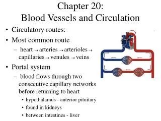
The Hepatic Portal System Carries blood from abdominal organs to liver Secondary capillary bed, organ Includes veins that drain blood from capillaries 9in the spleen, stomach, intestine and pancreas • Superior mesenteric vein • Splenic vein • Gastric, pancreatic, inferior mesenteric veins • Sinusoids
Circulation Physiology • Blood exchanges oxygen, carbon dioxide, other substances generated by cells • Tissue fluid (interstitial fluid) is exchange medium
Capillary Exchange How substances move between cells and capillary blood • Diffusion • Main process • Blood pressure • Moves material into tissue fluid • Osmotic pressure • Moves material into capillaries
The Dynamics of Blood Flow Vasomotor center in medulla regulates vasomotor activities • Vasodilation • Vasoconstriction • Precapillary sphincter • Widens to allow more blood to enter when tissues need more oxygen
Return of Blood to the Heart Mechanisms that promote blood’s return to heart • Contraction of skeletal muscles • Valves • Breathing • Chest expansion causes pressure to drop in the thorax, this action serves to push and pull blood through these cavities and return it to the heart
The Pulse • Use 2nd or 3rd finger to obtain • Ventricular contraction • Wave of increased pressure • Begins at heart and travels to arteries • Influenced by various factors • Body size • Gender • Age • Newborn-120-140 • Adult-60-100 • Muscular activity • Emotion • Body temperature • Thyroid secretion
Blood Pressure • Force exerted by blood against vessel walls • Determined by heart’s output and resistance to blood flow
Cardiac Output • Volume of blood pumped out of each ventricle in • one minute • Heart rate • Beats per minute • Stroke volume • Controlled by force of contractions
Resistance to Blood Flow Peripheral resistance is affected by • Vasomotor changes • Baroreceptors in large arteries • Elasticity of blood vessels • Viscosity • Total blood volume
Blood Pressure Measurement Pressure is measured in the brachial arm artery using a sphygmomanometer • Systolic pressure • Occurs during heart contraction • Normal systolic: 120 mmHg • Diastolic pressure • Occurs during heart relaxation • Normal diastolic: 80 mmHg
Figure 14-11 Blood pressure. In which vessels does the pulse pressure drop to zero?
Case Study Pathway of an Embolus From the Femoral Vein to the Pulmonary Artery Femoral vein External iliac vein Common iliac vein Inferior vena cava Right atrium Right ventricle Pulmonary trunk Pulmonary arteries