Download
1 / 43

E N D
The anterior teeth should not be positioned any further forward than the depth of the labial vestibule, determined by a line drawn from the depth of the vestibule and perpendicular to a line drawn parallel to the occlusal plane. The depth of the vestibule is the fulcrum point and the further forward a tooth extends beyond this point, the greater are the forces that tend to dislodge the denture.
The middle of the depth of the vestibule should be recorded on the maxillary & mandibular casts. The middle of the crest of the mandibular ridge should also be recorded. These are marked on the land areas because the baseplate will cover the marks on the ridge or in the vestibules.
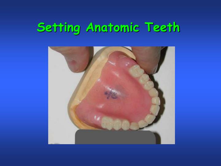
Next, mark the midline of the patient’s face by placing a dot on the incisive papilla and marking this midline on the maxillary anterior land area, extending down the front of the cast. The incisive papilla is a much more reliable landmark for the midline than the labial frenum.
Make a cut with a heated, sharp knife, at the midline in the anterior wax rim. Cut all the way to the baseplate. Make a similar cut just distal to the canine point. Remove this section of wax in its entirety. Reduce the anterior ridge of the maxillary baseplate to provide a little extra space for the tooth, but reduce the tooth if necessary to fit properly.
Use a flat plate resting flush with the occlusion rim to position the central incisor so that it contacts the occlusal plane. Set the rest of the anterior teeth on the right side according to the curve defined by the plastic ruler. The labioincisal line angle of the incisors should touch the ruler, as well as the midbuccal surface of the canine.
Use a flexible plastic ruler to verify that the incisal portion of the tooth’s labial surface is properly located and in contact with the anterior curvature of the occlusion rim.
An anterior view of the maxillary anterior teeth shows that only the lateral incisors do not touch the occlusal plane as recorded by mandibular wax rim. With the maxillary anterior teeth set, record the patient’s midline and set the mandibular anterior teeth.
The maxillary cast is placed back on the articulator. A quick look will show that the maxillary teeth are set on the same plane that the mandibular occlusion rim occupies, showing the value of having the maxillary and mandibular rims made in intimate contact. If the maxillary teeth are not on the same plane as the lower rim, they must be reset.
Mark the midline of the mandibular ridge on the mandibular wax rim and cut out a section representing the right mandibular anterior teeth from the rim. Reduce the thickness of the Triad baseplate from the middle of the ridge to the labial to allow for easier setting of the mandibular anterior teeth. A central incisor placed in this space shows that considerable reduction must be made on the cervico-lingual ridgelap surface before the tooth can be set in position.
Mark the midline of the mandibular ridge on the mandibular wax rim and cut out a section representing the right mandibular anterior teeth from the rim. Reduce the thickness of the Triad baseplate from the middle of the ridge to the labial to allow for easier setting of the mandibular anterior teeth. A central incisor placed in this space shows that considerable reduction must be made on the cervico-lingual ridgelap surface before the tooth can be set in position.
Remove enough wax to allow setting most of the posterior teeth and thin the baseplate to allow for their positioning. Leave a small segment of the wax rim intact and clearly mark the midline to facilitate the setting of the teeth anterior to it. After these teeth are set, remove this pillar to allow the last tooth to be set, and those teeth anterior to it will indicate the center of the ridge.
Check the position of the teeth to the center of the ridge with a tongue blade used as a straight edge. Once it is verified by your lab bench instructor that this relationship is correct, you may begin setting the teeth on the other side, using the same steps for setting, sequencing, and verification that were used on the other side.
With all the mandibular teeth set, check the occlusal plane with a flat metal plate to verify that all the teeth contact the flat surface evenly. Expect some changes because of the shrinkage of the wax, which will shift the teeth slightly. Correct any discrepancies before moving on to setting the maxillary posterior teeth.
With all the mandibular teeth set, check to see that all the teeth contact the flat surface of the maxillary wax rim evenly. If these teeth contact the wax rim evenly, begin setting the maxillary posterior teeth.
Remove the wax on one side of the maxillary baseplate. A quick look will verify that the maxillary posterior teeth should have plenty of space to be set with little or no reduction. The rim is left intact on the opposite side because this will help you to maintain the location of the occlusal plane.
Set the teeth on the maxillary right side so that the mesiolingual cusp of the maxillary first molar rests in the central fossa of the mandibular first molar. Set the teeth so that the buccal surfaces of the premolar(s) and mesial cusp of the first molar line up with the mid-buccal surface of the canine. The distobuccal cusp of the first molar should deviate approximately 20o from this plane and the second molar will fall along this plane.
If a maxillary second molar were set, the buccal cusps should line up with a second plane that is determined by the buccal surfaces of the maxillary first molar. In this case, there was not room enough to do so, because the opposing mandibular tooth would have to be set on the incline of the retromolar pad.
Set the teeth on the maxillary right side so that the mesiolingual cusp of the maxillary first molar rests in the central fossa of the mandibular first molar. Then move it up so that the distobuccal cusp is ½ mm off the flat plane. If there were room to set a maxillary second molar, its mesiobuccal cusp would be 1 mm off the flat plane and its distobuccal cusp would be 1½ mmoff the flat plane.
Adjust the occlusal surfaces of all posterior teeth to contact their opposing counterparts properly. Note: There is no vertical overlap of the anterior teeth at this time. With the anatomical maxillary posterior teeth properly positioned and having established a compensating curve and in harmonious centric occlusion with the anatomical mandibular posterior teeth, the mandibular anterior teeth are moved up to a point where the mandibular incisors contact the maxillary central incisors in protrusive and simultaneously with posterior teeth.
The mandibular molars are now adjusted so that they contact the maxillary molars in their new positions. The teeth are designed to fit together with the mandibular teeth to the outside of a curve. If they are set flat, there will be spaces between the maxillary molars. There should be a definite mediolateral component (Curve of Wilson in natural teeth) visible when looking from the back of the articulator .
The occlusal rims contoured to hold the lips in proper position. The teeth properly set in the wax to maintain desired labial fullness. The teeth set too far labially giving the lips a “pouched out”appearance. The teeth set too far lingually giving the lips a “dished in” appearance.
B C A Maximum horizontal overlap is shown in illustration B. If teeth are set as in A the mandibular denture would be very unstable during function. When vertical overlap is desired, the guide table must be set to record the anterior guidance.
The position of the natural central incisors and their relationship to the ridge.
A ridge immediately after the removal of the tooth. Red dotted line indicates the position of the natural root.
The direction of resorption in the maxillary anterior ridge is up and back. The solid line identifies the resorbed ridge. The red dotted line identifies the original contour of the ridge.
One of the most common errors in tooth positioning is positioning the teeth over the ridge without considering the original position of the natural teeth.
The denture with teeth set over the ridge is superimposed over the original position of the natural teeth. The loss of vertical dimension and lip support, and the inevitable resulting loss in aesthetics, is readily apparent.
The anterior teeth should not be positioned any further forward than the depth of the labial vestibule, determined by a line drawn from the depth of the vestibule and perpendicular to a line drawn parallel to the occlusal plane. The depth of the vestibule is the fulcrum point and the further forward a tooth extends beyond this point, the greater are the forces that tend to dislodge the denture.
Force increases with distance from the fulcrum. If the teeth are inclined anteriorly to compensate for a Class II relationship, a load placed anterior to the middle of the anterior vestibule will have the same destabilizing effect on the lower denture as the man on a seesaw.
HANAU’S QUINT Hanau described five factors (Hanau’s Quint) that affect occlusal balance • Condylar Inclination – We don’t change. • Incisal Guidance – Not with dentures • Occlusal Plane Inclination – Start with establishing plane parallel with Ala-Tragus line. • Cuspal Inclination – 0o, 10o, 20o, 30o, 33o • Compensating Curve – Set teeth (curve of Spee, curve of Wilson).
HANAU’S QUINT Condylar Inclination Compensating Curve Incisal Guidance – Not in dentures. Cuspal Inclination Occlusal Plane Inclination Cuspal inclination + Occlusal Plane Inclination Condylar inclination & Compensating Curve
Tooth Alignment • Centrals • Laterals • Canines
Maxillary Anterior Teeth The axial inclination of maxillary anterior teeth in the ‘basic’ arrangement. When viewed facially, all the anterior teeth are tilted mesially, with the lateral incisor inclined the most and raised about 1-2 mm above the plane.
Maxillary Anterior Teeth Cuspid Lateral Central When viewed laterally, the incisors are depressed at the cervical, with the lateral incisor being the most depressed, and the canine being straight with the long axis perpendicular to the occlusal plane.
Mandibular Anterior Teeth The axial inclination of mandibular anterior teeth in the ‘basic’ arrangement. When viewed facially, all the anterior teeth except the central incisors are tilted mesially, with the canine inclined the most.
Mandibular Anterior Teeth Cuspid Lateral Central When viewed laterally, the mandibular central incisors are depressed at the cervical, the lateral incisor is straight, and the canine is inclined lingual to the long axis.
Antero-posterior positioning of anterior teeth give support to the lips, cheeks, and other tissues of the oral cavity. The replacement of artificial teeth in the original position of the natural teeth frequently is not stressed or simply overlooked and resorbed residual ridges are used as the primary control of tooth position. A resorbed residual ridge – because of what may be extreme changes in shape and size – is a questionable landmark for either functional or esthetic tooth position.
Three different approaches to providing a denture on the same patient. Note the differences in the setting of the teeth and the festooning. The same mold of teeth was used in each case.
? ? ? ? ? ? ? Questions? Questions? Questions?