Download
1 / 19

E N D
When viewed facially, all maxillary anterior teeth are tilted mesially, with the lateral incisor inclined the most and raised about 1-2 mm above the plane. When viewed laterally, the incisors are depressed at the cervical, with the lateral incisor being the most depressed, and the canine being straight with the long axis perpendicular to the occlusal plane.
When viewed facially, all the mandibular anterior teeth except the central incisors are tilted mesially, with the canine inclined the most. When viewed laterally, the central incisors are depressed at the cervical, with the lateral incisor being straight, and the canine being inclined lingual to the long axis.
The anterior teeth should not be positioned any further forward than the depth of the labial vestibule, determined by a line drawn from the depth of the vestibule and perpendicular to a line drawn parallel to the occlusal plane. The depth of the vestibule is the fulcrum point and the further forward a tooth extends beyond this point, the greater are the forces that tend to dislodge the denture.
The middle of the depth of the vestibule should be marked on the land areas of the maxillary & mandibular casts. Next, mark the midline of the patient’s face by placing a dot on the incisive papilla and marking this midline on the maxillary anterior land area, extending down the front of the cast.
Mark the midline of the palate on the anterior wax rim using the middle of the incisive papilla as a guideline.
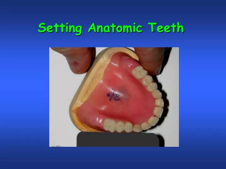
Make a cut with a heated, sharp knife, at the midline in the anterior wax rim. Cut all the way to the baseplate. Make a similar cut just distal to the canine point. Remove this section of wax in its entirety. Reduce the anterior ridge of the maxillary baseplate to provide a little extra space for the tooth, but reduce the tooth if necessary to fit properly.
Melt the wax in the area where the section was removed with a hot instrument. Set the maxillary central incisor, being careful to not disturb the midline as recorded by the remaining section of wax rim. Check its position with a flat plate.
Note that the central incisor contacts the flat plate resting on the occlusal plane. Set the rest of the anterior teeth on the right side according to the curve defined by the plastic ruler. The labioincisal line angle of the incisors should touch the ruler, as well as the midbuccal surface of the canine.
Note that the lateral incisor is set about 1mm above the occlusal plane.
…then check to make sure the alignment is correct with the contour of the wax rim, using a flexible ruler. Set the teeth on the right side following the same procedures.
An anterior view of the maxillary anterior teeth shows that only the lateral incisors do not touch the occlusal plane as recorded by mandibular wax rim. With the maxillary anterior teeth set, the patient’s midline is recorded and proper labial support is established.
Note: If the patient has an Angle’s Class II musculoskeletal relationship (Overjet) they may want you to set their teeth further back on the ridge. If you do this, they will ask you to remove the denture flange, because they feel this is too thick and pushes the lip out. You cannot do this because you would decrease the denture bearing area and lose the vacuum seal you have under the maxillary denture. If you make the flange too thin, it will break and the patient will not want it thickened in the repair. Avoid this situation by properly educating the patient on their Class II relationship prior to making the denture.
The maxillary cast is placed back on the articulator. A quick look will show that the maxillary teeth are set on the same plane that the mandibular occlusion rim occupies. If the maxillary teeth are not on the same plane as the lower rim, they must be reset.
Record the midline on the mandibular wax rim and follow the same procedures used on the maxillary anterior teeth to set the mandibular anterior teeth. Cut out a section representing the right mandibular anterior teeth from the rim.
Reduce the thickness of the Triad baseplate from the middle of the ridge to the labial to allow for easier setting of the mandibular anterior teeth. A central incisor placed in this space shows that considerable reduction must be made on the cervico-lingual ridgelap surface before the tooth can be set in position.
Set the mandibular anterior teeth using the same sequencing and procedures as were used with the maxillary anterior teeth. Note that all the mandibular anterior teeth contact the occlusal plane in centric relation.
Review of steps • Mark midline on max. rim. • Cut this midline through to the baseplate. • Cut a line distal to the canine on one side. • Remove this entire section. • Set the central incisor, then the lateral, then the canine. • Check the height with a flat plate. • Check the labial position with a flexible ruler. • Cut out a section of the wax rim from the midline to the distal of the other canine. • Repeat steps 4-7 with these teeth. • Mark the midline on the mandibular rim using the maxillary midline as a reference. • Remove a section of the wax rim from the midline to the distal of a canine. • Complete the same sequence of steps on the lower anterior teeth as was done on the maxillary teeth.
? ? ? ? ? ? ? Questions? Questions? Questions?