Download
1 / 43
430 likes | 469 Views
Paediatric patient dose surveys. Colin Martin and David Sutton. Overview. Introduction 1. Chest and abdominal radiographs 2. Dynamic procedures. Why are kids special?. higher risk than for adults often different techniques less data available

E N D
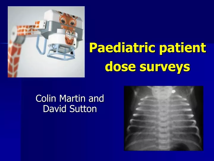
Paediatric patient dose surveys Colin Martin and David Sutton
Overview Introduction 1. Chest and abdominal radiographs 2. Dynamic procedures
Why are kids special? higher risk than for adults often different techniques less data available only performed occasionally in some hospitals lack of optimisation
Radiation risk in children Greater chance for expression of radiation induced effects Greater sensitivity for some cancers High frequency for some examinations Lack of cooperation and optimisation
Large numbers of images can be taken of new born babies K. Smans, Leuven
Sometimes over 100 images can be taken of new borns C. Maccia, Paris
Some Paediatric Exams Water soluble contrast studies Mict Cystogram for reflux Intussusception Non-accidental injury assessment Hips for displasia
Fewer patients • Paediatric exams only represent 1-2% of those in UK database (despite extra efforts to collect data) • Only paediatric hospitals have sufficient patients for full survey • Other hospitals may have only a handful of patients even for commonest exams
X-ray equipment for paediatrics Generators Filtration (additional copper) Paediatric anti-scatter grids Automatic exposure control Low dose fluoroscopy KAP meters with resolution of 0.1 mGy cm2 The Ideal is to use dedicated equipment. Factors to consider include:
1. Paediatric Radiography • Simple examinations • Assess ESD or KAP • Large range in patient size
Best Practice Guidelines High speed screen/film systems Avoid use of anti-scatter grids where possible Additional filtration - copper High kV-short exposure techniques Gonad protection Dedicated equipment Trained staff Cook et al, 1998
Use of Grids • Use of an anti-scatter grid can double the dose • Children’s bodies are small • so there is less scattered radiation • grids are often unnecessary
New born child can be placed directly on cassette for radiograph
Influence of grid on doseEffect of introduction of grid for larger children
Good collimation for neonatesAchieved by positioning lead/rubber drapes on incubator Poor radiation protection Good radiation protection
Copper filtration:Removes low energy photons that are absorbed in superficial tissues Spectra of 80 kV X-ray beams with and without 0.2 mm copper X-ray photons removed Reduces skin dose Increase in mAs required Maintains good contrast Exposure adjusted to give similar dose at the image receptor
Dose quantities Radiography ESD : measured with TLD or calculated from tube output KAP : If KAP meter fitted Fluoroscopy KAP : measured with KAP meter
Dosimetry Problems low readings (KAP meters/TLD) give poor precision TLD may be visible or cause practical difficulties calculations of dose complicated by variable patient size small sample sizes uncertainties in effective dose and risk factors
Exposure parameters & tube output measurements Need tube output (as a function of kV) from the medical physics report Need kV, distance and mAs values for exposure Uncertainties: Accuracy of the distance data Field size
Paediatric Data Collection Exam Patient data essential age Height & weight thickness Focus to Skin Distance Use of grid Filtration
Why is size a problem ? Continuous size distribution (neonate to adult) Establishment of DRLs and comparisons with reference levels need to be meaningful Conflict arises between sample size and variability arising from patient size
Increase in KAP with patient sizeNote that vertical axis is a log scale Courtesy T. Kiljunen, STUK, Finland
Possible Approaches Age banding Patients grouped together to provide sufficient data Retrospective data analysis Correction of data using effective attenuation coefficients
Size correction using effective attenuation coefficients Corrections can be made to data to enable patients with a range of ages/sizes to be used Equate to a patient of standard thickness ‘s’ FESD = eµ(s-d) Where = effective attenuation coefficient (derived from measurements of entrance and exit doses) with varying phantom thicknesses – fixed kV d = thickness of patient for which data collected Hart et al (2000): NRPB-R318
h Equivalent cylinder diameter • Patient diameter can be equated to a standard cylinder of the same length and weight, if other data not available • Equivalent diameter = 2√[Weight/( Height)] Density = ( 1 g cm3)
Size correction for KAP Similar corrections can be made to KAP data FKAP = eµ(s-d)s2/d2 = effective attenuation coefficient S = thickness of standard patient d = thickness of patient for which data collected Incorporates FSD correction Field size correction for KAP Hart et al (2000): NRPB-R318
Age ranges • 0 – 6 months newborn • 7 m – 23 m 1 y old infant • 2 – 7 y 5 y old • 8 y – 12 y 10 y old
National patient dose survey • Collect data from hospitals • Input data into database • Calculate mean KAP or ESD • Compare data from all centres
National SurveyDistribution of doses for paediatric chest • Identify the 3rd quartile dose for use as DRL • 75% below DRL; 25% above need optimisation 3rd Quartile - 70 mGy
Setting DRL based on ESD data for agreed age band Third Quartile DRL 70 mGy
Regional patient dose survey • Compare data from all centres • For non-paediatric hospitals it may be necessary just to collect standard factor data for some examinations • Investigate possible reasons for high doses above DRL • Produce individual report for each centre • Liaison with individual hospitals is necessary to ensure optimisation of protection
Issues with survey data … • Include patient weight • 20+ patients if possible • Reset KAP meter after each examination • Quote KAP in correct units • Often large differences between centres
National DRL Neonatal dose data from factors used in District General Hospitals • Hospitals rarely carry out examinations and factors are not optimised
Reasons for high DAP values- Radiographic • Low patient numbers • Patient weight not given • Using a low kV • KAP quoted in wrong units • KAP meter not reset after each patient/examination • Different techniques used
2. Dynamic examinations Less frequent KAP measurements Effect of the weight? More complicated to calculate effective doses
DAP v age for MCU BJR 72:763-772 (1999) Courtesy of F. Schultz
Reasons for high DAP values • Patient weight not given • Low patient numbers • High pulse rate • Old equipment • Large variation in screening times • Protocols not being followed
Summary Paediatric dosimetry is not straightforward Dose measurement/calculation methodologies need to be well thought out A consistent approach to patient size correction is needed Dose quantities need to be appropriate for task