Download
1 / 28
280 likes | 420 Views
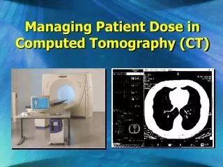
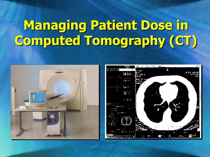
Managing Patient Dose in Computed Tomography (CT). International Commission on Radiological Protection. Information abstracted from ICRP Publication 87 Available at www.icrp.org

E N D
International Commission on Radiological Protection • Information abstracted from • ICRP Publication 87 • Available at www.icrp.org • Task Group: M.M. Rehani, G. Bongartz, S.J. Golding, L.Gordon, W. Kalender, T. Murakami, P. Shrimpton, R. Albrecht, K. Wei
Use and Disclaimer • This is a PowerPoint file • It may be downloaded free of charge • It is intended for teaching and not for commercial purposes • This slide set is intended to be used with the complete text provided in ICRP Publication 87
Contents • Situation analysis • Why increased frequency? • Why increased dose? • Is the dose really high? How high? • What can be done to manage patient dose? • What can operator do? • Action for manufacturer • Action for physician & radiologist
Situation Analysis • CT continues to evolve rapidly despite many advances in other imaging modalities • It is one of the most important radiological examinations worldwide • The frequency of CT examinations is increasing rapidly from 2% of all radiological examinations in some countries a decade ago to 10-15 % now • Patient doses in CT have not decreased in contrast to radiography where nearly 30% reduction has been documented in last decade
What is the dose from CT? How high? • The effective dose in chest CT is in the order of 8 mSv (around 400 times more than chest radiograph dose) and in some CT examinations like that of pelvic region, it may be around 20 mSv (1000 CXRs) • The absorbed dose to tissues from CT can often approach or exceed the levels known to increase the probability of cancer as shown in epidemiological studies
Radiation Exposure by Source/Test Chest X-Ray 0.02 mSv (1) Lumbar Spine 1.3 mSv (65) CT Chest 8 mSv (400) CT Abdomen or Pelvis 10 mSv (500) Virtual Colonoscopy 36-50 mSv(720-1000) Hiroshima Survivors 50-200mSv
A. Contributionsto frequency B. Contributionsto prescribed dose 15% 41% 12% 8% 9% 2% 5% 1% 1% 6% 29% 10% 34% 5% UNSCEAR 2000
Why Increased Frequency? • 20 years ago, a standard CT of the thorax took several minutes while today similar information can be accumulated in a single breath hold making it attractive, patient & user friendly • Advances in CT technology have made possible CT fluoroscopy and interventional procedures, in some cases replacing ultrasound guided interventions • Repeat CT exams
Why increased dose… • Unlike radiography where over-exposure results in blackening of film, better image quality is obtained with higher exposures in CT • There is a tendency to increase the volume covered in a particular examination • Modern helical CT involves volume scanning with no inter-slice gap and with possibility of overlapping scans • Repeat CT examinations
Why increased dose (cont’d) • Same exposure factors used for children as for adult • Same exposure factors for pelvic (high contrast region) as for abdomen (low contrast region)
Organ doses in CT • Breast dose in thorax CT may be as much as 30-50 mGy, even though breasts are not the target of imaging procedure • Eye lens dose in brain CT, thyroid in brain or in thorax CT and gonads in pelvic CT receive high doses
Tissues in the field although they are not the area of interest for the procedure Lens of the eye Breast tissue
Cataract in eye of interventionist after repeated use of over table x-ray tube
Informed consent and records • Patients are entitled to know the risks of radiation injury if likely to be high • A written record should be kept if skin doses are estimated to be >3 Gy (1 Gy for repeated procedures) • Not all skin reactions are due to contrast medium allergy
Atrophic indurated plaque Hyper & hypo pigmentation, with telangiectasia Chronic radiodermatitis in 17 year old female patient after x2 radiofrequency ablation procedures
Follow-up • Radiation skin injury may present late and the association not considered if no documentation • All patients with estimated skin doses of 3 Gy should be followed up 10-14 days after exposure • A system to identify repeat procedures should be set up
Example of chronic skin injury due to cumulative skin dose of ~20,000 mGy (20 Gy) from coronary angiography and x2 angioplasties 21 months after first procedure, base of ulcer exposes spinous process
Leukemia and Cancer • Most interventional procedures are performed on older patients where benefit almost always outweighs radiation risk • The radiation risk increases progressively with younger age groups • Radiation has been shown to increase the risk for leukemia and many types of cancer in adults and children
Does spiral CT give more or less radiation dose? • It depends upon the choice of factors • Even though it is possible to perform a spiral CT with lower radiation dose than slice-by-slice CT, in practice the patient gets higher dose due to the factors chosen (scan volume, mAs, pitch, slice width
Does multi-slice CT impart more or less radiation dose? • An increase by 10-30% may occur with multi-slice detector array
Some Observations • Most doctors including many radiologists have a feeling that modern CT scanners which are very fast give lesser radiation dose • Unfortunately ‘time’ and ‘radiation dose’ are not proportional in such a situation • Over the years the x-ray tubes are becoming more and more powerful such that they can give high bursts of x-rays which can give satisfactory image in shorter exposure time
Actions for Physician & Radiologist… • Justification: Ensure that patients are not irradiated unjustifiably • Request for CT examination should be generated only by properly qualified medical or dental practitioners depending upon national educational and qualification system. The physician is responsible for weighing the benefits against risks • Clinical guidelines advising which examinations are appropriate and acceptable should be available to clinicians and radiologists
Actions for Physician & Radiologist (cont’d) • Consider whether the required information be obtained by MRI, ultrasonography • Consider value of contrast medium enhancement prior to commencing examination • CT scanning in pregnancy may not be contraindicated, particularly in emergency situations, although examinations of the abdomen or pelvis should be carefully justified
Actions for Physician & Radiologist (cont’d) • CT examination shouldnot be repeatedwithout clinical justification and should be limited to the area of interest • Clinician has theresponsibilityto communicate to the radiologist about previous CT examination of the patient • CT examination forresearchpurposes that do not have clinical justification (immediate benefit to the person undergoing the examination) should be subject to critical evaluation by an ethics committee
Actions for Physician & Radiologist (cont’d) • CT examination of chest in young girls and young females needs to be justified in view of high breast dose • Once the examination has been justified, radiologist has the primary responsibility for ensuring that the examination is carried out with good technique
Web sites for additional information on radiation sources and effects • European Commission(radiological protection pages):europa.eu.int/comm/environment/radprot • International Atomic Energy Agency:www.iaea.org • International Commission on Radiological Protection:www.icrp.org • United Nations Scientific Committee on the Effects of Atomic Radiation:www.unscear.org • World Health Organization:www.who.int