Download
1 / 77
830 likes | 1.24k Views
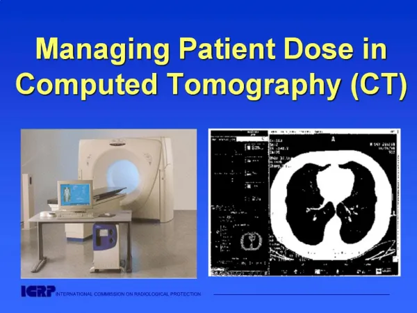
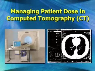
Patient Dose Management. L 5b. Factors that influence Patient Absorbed Dose. Procedural-related factors Positioning of image receptor and X ray source relative to the patient Beam orientation and movement Collimation Acquisition and fluoroscopic technique factors on some units

E N D
Factors that influence Patient Absorbed Dose • Procedural-related factors • Positioning of image receptor and X ray source relative to the patient • Beam orientation and movement • Collimation • Acquisition and fluoroscopic technique factors on some units • Fluoroscopy pulse rate • Acquisition frame rate • Total fluoroscopy/acquisition time Lecture 5: Patient Dose Management
Positioning of image receptorand X ray source relative to the patient
Only a small percentage (typically ~1%) penetrate through to create the image. Beam entering patient typically ~100x more intense than exit beam in average size patient Lecture 5: Patient Dose Management
Inverse Square Law X ray intensity decreases rapidly with distance from source; conversely, intensity increases rapidly with closer distances to source. 1 unit of intensity 4 units of intensity 16 units of intensity 64 units of intensity 8.8 cm 17.5 cm 35 cm 70 cm Lecture 5: Patient Dose Management
Automatic Brightness Control (ABC) Image Handling and Display Image Receptor Automatic Dose Rate Control Operator Patients Electrical Stabilizer Foot Switch X ray tube Operator Controls Power Controller High-voltage transformer Primary Controls Feedback circuitry from the image receptor communicates with the X ray generator modulates X ray output to achieve appropriate subject penetration by the X ray beam and image brightness. Lecture 5: Patient Dose Management
2 units of intensity Image Receptor Inverse Square Law (1) All other conditions unchanged, moving image receptor toward patient lowers radiation output rate and lowers skin dose rate. 4 units of intensity Image Receptor Image Receptor Lecture 5: Patient Dose Management
2 units of intensity Image Receptor Inverse Square Law (1) 4 units of intensity Image Receptor Image Receptor Lesson: Keep the image intensifier as close to the patient as is practicable for the procedure. Lecture 5: Patient Dose Management
Distance between patient and detector Lecture 5: Patient Dose Management
2 units of intensity 4 units of intensity 16 units of intensity 64 units of intensity Inverse Square Law (2) All other conditions unchanged, moving patient toward or away from the X ray tube can significantly affect dose rate to the skin Lesson: Keep the X ray tube at the practicable maximum distance from the patient. Lecture 5: Patient Dose Management
Distance between patient and X ray source Lecture 5: Patient Dose Management
Tall vs. Short Operators - Impact on Patient Dose? Lecture 5: Patient Dose Management
ISOCENTER Positioning anatomy of interest at the isocenter permits easy reorientation of the C-arm. This usually shortens the distance between the X ray tube and the patient, increasing the patient’s entrance port skin dose. Lecture 5: Patient Dose Management
ISOCENTER When isocenter technique is employed, move the image intensifier as close to the patient as practicable to limit dose rate to the entrance skin surface. Lecture 5: Patient Dose Management
Beam Orientation Physical factors and challenges to radiation management Lesson: Reorienting the beam distributes dose to other skin sites and reduces risk to single skin site. This is especially important in coronary angioplasty for chronic total occlusion. Lecture 5: Patient Dose Management
Overlap Areas in Beam Re-orientation Lesson: Reorienting the beam in small increments may leave area of overlap in beam projections, resulting in large accumulations for overlap area (red area). Good collimation can reduce this effect. Reproduced with permission from Wagner LK, Houston, TX 2004. Lecture 5: Patient Dose Management
Beam Orientation Physical factors and challenges to radiation management Conclusion: Orientation of beam is usually determined and fixed by clinical need. When practical, reorientation of the beam to a new skin site can lessen risk to skin. Overlapping areas remaining after reorientation are still at high risk. Good collimation reduces the overlap area. Lecture 5: Patient Dose Management
Imaging modes –Fluoroscopy, (Cine) Acquisition,Digital Subtraction Angiography
Fluoroscopy vs Cine Acquisition Influence of operation modes:from low fluoroscopy to cine, radiation / scatter dose rate could increase in a factor of 10-15 Lecture 5: Patient Dose Management
Can you tell ………. Which image is FLUOROSCOPY ? Which one is ACQUISITION?
Better image quality with higher radiation dose reaching the image receptor. Tradeoff: higher patient dose!! Image Quality Radiation Dose Lecture 5: Patient Dose Management
ALARA No known safe limit of magnitude of radiation exposure. As Low As Reasonably Achievable Physicians Patients Professional staff Lecture 5: Patient Dose Management
Siemens Axiom Artis, Fluoro low dose 20 cm PMMA 13 Gy/fr (entrance PMMA) Siemens Axiom Artis Cine normal mode 20 cm PMMA 177 Gy/fr (entrance PMMA) Lecture 5: Patient Dose Management
Lowest input dose needed to generate a USABLE image Set the default fluoroscopy mode to LOW Lecture 5: Patient Dose Management
Duration of Fluoroscopy/Cine Acquisition Influence of operation modes:from low fluoroscopy to cine, radiation / scatter dose rate could increase in a factor of 10-15 Important to keep in mindDURATIONof fluoroscopy fluoroscopy x 10-15 sec ~ cine x 1 sec Lecture 5: Patient Dose Management
Digital Image Subtraction (DSA) • Obtained by subtracting one image from another electronically removes information that is identical in 2 images • Subtraction process accentuates image noise counter this effect by acquiring each of the original images at a substantially (up to 20x) higher dose per frame. • Generally, studies that use DSA employ larger aggregate doses than do studies that employ unsubtracted cinefluorography. Lecture 5: Patient Dose Management
Design of fluoroscopic equipment for proper radiation control Pulsed Fluoroscopy Understanding Variable Pulsed Fluoroscopy Background: dynamic imaging captures many still images every second and displays these still-frame images in real-time succession to produce the perception of motion. How these images are captured and displayed can be manipulated to manage both dose rate to the patient and dynamic image quality.Standard imaging captures and displays 25 - 30 images per second. Lecture 5: Patient Dose Management
Each angiographic ‘run’ consists of multiple still images taken in quick succession. Lecture 5: Patient Dose Management [ video clip]
Continuous fluoroscopy In conventional continuous-beam fluoroscopy there is an inherent blurred appearance of motion because the exposure time of each image lasts the full 1/30th of a second at 30 frames per second. Images 30 images in 1 second X rays Continuous stream of X rays produces blurred images in each frame Lecture 5: Patient Dose Management
Pulsed fluoroscopy, no dose reduction Pulsed fluoroscopy produces sharp appearance of motion because each of 30 images per second is captured in a pulse or snapshot (e.g., 1/100th of a second). Images 30 images in 1 second X rays Each X ray pulse shown above has greater intensity than continuous mode, but lasts for only 1/100th of a second; no X rays are emitted between pulses; dose to patient is same as that with continuous fluoroscopy Lecture 5: Patient Dose Management
Pulsed Fluoroscopy Fluoroscopic pulsing X rays are produced during a small portion of the video frame time. The narrower the pulse width, the sharper the image. ( “Faster shutter speed” in camera ) Lecture 5: Patient Dose Management
Pulsed Fluoroscopy Physical factors and challenges to radiation management Pulsed imaging controls: Displaying 25–30 picture frames per second is usually adequate for the transition from frame to frame to appear smooth. This is important for entertainment purposes, but not necessarily required for medical procedures. Manipulation of frame rate can be used to produce enormous savings in dose accumulation. Lecture 5: Patient Dose Management
Pulsed fluoroscopy, dose reduction at 15 pulses per second Sharp appearance of motion captured at 15 images per second in pulsed mode. Dose per pulse is same, but only half as many pulses are used, thus dose is reduced by 50%. The tradeoff is a slightly choppy appearance in motion since only half as many images are shown per second Images X rays 15 images in 1 second Lecture 5: Patient Dose Management
Pulsed fluoroscopy, dose reduction at 7.5 pulses per second Pulsed fluoroscopy at 7.5 images per second with only 25% the dose Images X rays Average 7.5 images in 1 second Lecture 5: Patient Dose Management
Pulsed fluoroscopy, dose enhancement at 15 pulses per second Dose per pulse is enhanced because pulse intensity and duration is increased. Overall dose is enhanced. Images Images X rays 15 images in 1 second X rays 15 images in 1 second Reproduced with permission from Wagner LK, Houston, TX 2004. Lecture 5: Patient Dose Management
Variable Pulsed Fluoroscopy Design of fluoroscopic equipment for proper radiation control Lesson: Variable pulsed fluoroscopy is an important tool to manage radiation dose to patients but the actual effect on dose can be to enhance, decrease or maintain dose levels. The actual effect must be estimated by a qualified physicist so that variable pulsed fluoroscopy can be properly employed. Lecture 5: Patient Dose Management
Collimation Lecture 5: Patient Dose Management
A word about collimation What does collimation do? Collimation confines the X ray beam to an area of the user’s choice. Lecture 5: Patient Dose Management
Collimation Why is narrowing the field-of-view beneficial? • Reduces stochastic risk to patient by reducing volume of tissue at risk • Reduces scatter radiation at image receptor to improve image contrast • Reduces scatter radiation to in-room personnel • Reduces potential overlap of fields when beam is reoriented Lecture 5: Patient Dose Management
Scattered Radiation Scattered radiation X-Ray • Two undesirable effects: • (1) predominant source of radiation exposure to the laboratory personnel; Lab Personnel Patient Operator Lecture 5: Patient Dose Management
Two undesirable effects: (2) scattered radiation that continues in the forward direction and reaches the image receptor decreases the quality (contrast) of the image Scattered Radiation Reduction of Image Contrast by Scattered Radiation Lecture 5: Patient Dose Management
Collimation: Contrast Improvement by Reducing X ray Beam Size Lecture 5: Patient Dose Management
Beam Orientation, Overlap and Collimation Lesson: Reorienting the beam in small increments may leave area of overlap in beam projections, resulting in large accumulations for overlap area (red area). Good collimation can reduce this effect. Lecture 5: Patient Dose Management
Collimation What collimation does NOT do – It does NOT reduce dose to the exposed portion of patient’s skin In fact, dose at the skin entrance site increases, sometimes by a factor of 50% or so, depending on conditions. Lecture 5: Patient Dose Management
Equipment-related factors Movement capabilities of C-arm, X ray source, image receptor Field-of-view size Collimator position Beam filtration Fluoroscopy pulse rate and acquisition frame rate Fluoroscopy and acquisition input dose rates Automatic dose-rate control including beam energy management options X ray photon energy spectra Software image filters Preventive maintenance and calibration Quality control Factors that influence Patient Absorbed Dose Lecture 5: Patient Dose Management
Image receptor degrades with time Image Handling and Display Image Receptor Automatic Dose Rate Control Operator Patients Electrical Stabilizer Foot Switch X ray tube Operator Controls Power Controller High-voltage transformer Primary Controls Lecture 5: Patient Dose Management