Download
1 / 27
270 likes | 521 Views
Anatomy and Embryology of the Pharynx. Jared Bradley Turner, MD January 7, 2005. Embryology. Components of branchial/pharyngeal apparatus: Pharyngeal arches Pharyngeal pouches Pharyngeal clefts/grooves. Pharyngeal (branchial) arches. Derived from neural crest cells

E N D
Anatomy and Embryology of the Pharynx Jared Bradley Turner, MD January 7, 2005
Embryology Components of branchial/pharyngeal apparatus: • Pharyngeal arches • Pharyngeal pouches • Pharyngeal clefts/grooves
Pharyngeal (branchial) arches • Derived from neural crest cells • Resemble fish gills (branchia) • Begin to develop early in the 4th week • By end of 4th week, four pairs of arches are visible on the surface (not 5th and 6th ) and a buccopharyngeal membrane ruptures forming communication between primitive oral cavity and foregut
Pharyngeal arches (cont.) • Contribute to the formation of the neck as well as the face. • Visible structures at 42 weeks: 1st arch: mandibular prominence, maxillary prominences, and the frontonasal prominence
Pharyngeal arches (cont.) • Core of mesenchymal tissue covered by surface ectoderm (outside) and by endodermal epithelium (inside) • Ectoderm -> skeletal • Mesoderm -> muscles with accompanying nerve • Arterial component (aortic arches) • Therefore, each arch carries nerve, muscle, bone and blood supply
First pharyngeal arch • Maxillary process (dorsal) • Premaxilla, maxilla, zygomatic bone, portion of temporal bone • Mandibular process (ventral) • Contains Meckel’s cartilage which disappears except for dorsal end (incus & malleus) and mandible
First pharyngeal arch • Muscles of mastication, digastric (ant belly), mylohyoid, tensor tympani and tensor palatini • Therefore, the accompanying motor nerve is the mandibular branch of trigeminal (V2) and sensory are V1, V2, and V3 • 1st aortic arch practically disappears but forms the maxillary artery
Second pharyngeal arch • Reichert’s cartilage – stapes, styloid process, stylohyoid ligament, lesser horn and upper part of the hyoid • Muscles include: stapedius, stylohyoid, digastric (post belly), auricular, and those of facial expression • Facial nerve (CN VII) • 2nd aortic arch – stapedial & hyoid arteries
Third pharyngeal arch • Cartilaginous contributions include greater horn and lower part of hyoid • Sole muscle: stylopharyngeus • CN IX (Glossopharyngeal nerve) • 3rd aortic arch (quite large): common carotid, 1st portion of internal carotid (remainder dorsal aorta), and external carotid
Fourth & sixth pharyngeal arch • Cartilaginous contributions to larynx derived from fusion: thyroid, cricoid, arytenoid, corniculate, and cuneiform • Muscles of 4th: cricothyroid, levator palatini, and pharyngeal constrictors are innervated by SLN (CN X) • Muscles of 6th: intrinsics of larynx are innervated by RLN (CN X) • 4th aortic arch: L->arch of aorta & R->subclavian • 6th aortic arch: L & R pulmonary with ductus arteriosus on left
Pharyngeal pouches (5) • 1st:tubotympanic recess-> middle ear & eustacian tube -> TM • 2nd palatine tonsil/fossa • 3rd: inferior parathyroid (dorsal), thymus (ventral) • 4th: superior parathyroid • 5th: ultimobranchial body -> calcitonin producing C cells (parafollicular)
Pharyngeal clefts/grooves (4) • 1st: external auditory meatus • 2nd-4th : epicardial ridge and cervical sinus (disappears)
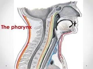
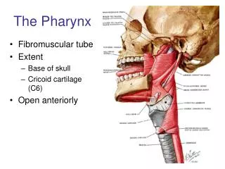
Anatomy (cont.) • Extends from base of skull to inferior border of cricoid cartilage anteriorly and inferior border of C6 posteriorly • Widest portion (5cm) at hyoid • Narrowest portion (1.5cm) at caudal end • Divided into 3 parts: nasopharynx, oropharynx, and laryngo(hypo)pharynx
Nasopharynx • Respiratory function • Anterior: choana (posterior nasal aperture) • Posterior: pharyngobasilar membrane and superior constrictor muscle • Superior: basilar portion of occipital bone • Inferior: soft palate
Oropharynx • Digestive function • Anterior: anterior tonsillar pillar • Posterior: superior constrictor • Superior: soft palate • Inferior: base of tongue, superior epiglottis • Laterally: palatoglossal and palatopharyngeal arches
Hypopharynx • Lies posterior to the larynx • Superior: superior border of epiglottis and pharyngoepiglottic folds • Inferior: inferior border of the cricoid • Posterior/lateral: middle & inferior constrictors, bodies of C4-C6 • Anterior: laryngeal inlet
Pharyngeal muscles • External circular and internal longitudinal (opposite in remainder of GI tract) • External: 3 constrictors (CN XI via X and ELN/RLN for middle and inferior) function to constrict wall of pharynx during swallow • Internal: palatopharyngeus and salpingopharyngeus (CN XI via X) and stylopharyngeus (CN IX) act to elevate pharynx and larynx during speech/swallow
Pharyngeal muscles • Tensor veli palatini (V3) tenses soft palate & opens ET during yawn/swallow • Levator veli palatini (CN XI via X) elevates palate during swallow/yawn • Palatoglossus (CN XI via X) approximates tongue and soft palate
Oral cavity I, II, III Oro/hypopharynx deep II, III, IV Nasopharynx II, V, III Pharyngeal lymphatic drainage