Download
1 / 43
470 likes | 882 Views
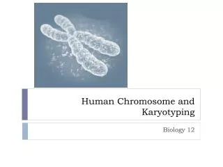
Chapter 5 Human Chromosomes and Chromosome Behavior. Human Chromosomes. Humans contain 46 chromosomes, including 22 pairs of homologous autosomes and two sex chromosomes

E N D
Chapter 5 Human Chromosomes and Chromosome Behavior
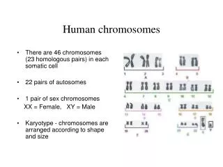
Human Chromosomes • Humans contain 46 chromosomes, including 22 pairs of homologous autosomes and two sex chromosomes • Karyotype = stained and photographed preparation of metaphase chromosomes arranged according to their size and position of centromeres
Figure 5.1A: Human chromosome painting Figure 5.1B: Human chromosome painting Parts A and B Courtesy of Johannes Wienberg, Ludwig-Maximillians-University, and Thomas Ried, National Institutes of Health
Centromeres • Chromosomes are classified according to the relative position of theircentromeres • Inmetacentric it is located in middle of chromosome • Insubmetacentric — closer to one end of chromosome • Inacrocentric — near one end of chromosome • Chromosomes with no centromere, or with two centromeres, are genetically unstable
Human Chromosomes • Each chromosome inkaryotypeisdivided into two regions (arms) separated by the centromere • p = short arm (petit); q= long arm • p and q arms are divided into numbered bands and interband regions based on pattern of staining • Within each arm the regions are numbered.
Figure 5.3: Designations of the bands and interbands in the human karyotype.
Human X Chromosome • Females have two copies of X chromosome • One copy of X is randomly inactivated in all somatic cells. • Females are genetic mosaics for genes on the X chromosome; only one X allele is active in each cell. • Barr body = inactive X chromosome in the nucleus of interphase cells. • Dosage compensationequalizes the number of active copies of X-linked genes in females and males.
Figure 5.7: Schematic diagram of somatic cells of a normal female
The calico cat shows visible evidence of X-chromosome inactivation. Figure 5.8: Female cat heterozygous for the orange and black coat color alleles
Human Y Chromosome • Y chromosome is largely heterochromatic. • Heterochromatin is condensed inactive chromatin. • Important regions of Y chromosome: • pseudoautosomal region: region of shared X-Y homology • SRY – master sex controller gene that encodes testis determining factor (TDF) for male development
The pseudoautosomal region of the X and Y chromosomes has gotten progressively shorter in evolutionary time. Figure 5.9: Progressive shortening of the mammalian X-Y pseudoautosomal region through time Data from B.T. Lahn and D.C. Page, Science 286 (1999): 964-967
Human Y Chromosome • Y chromosome does not undergo recombination along most of its length, genetic markers in the Y are completely linked and remain together as the chromosome is transmitted from generation to generation • The set of alleles at two or more loci present in a particular chromosome is called a haplotype • The history of human populations can be traced through studies of the Y chromosome
Abnormal Chromosome Number • Euploid= balanced chromosome abnormality = the same relative gene dosage as in diploids (example: tetraploids) • Aneuploid= unbalanced set of chromosomes = relative gene dosage is upset (example: trisomy of chromosome 21) • Monosomic= loss of a single chromosome copy Polysomic= extra copies of single chromosomes • Chromosome abnormalities are frequent in spontaneous abortions.
Abnormal Chromosome Number • Monosomy or trisomy of most human autosomes is unviable. There are three exceptions: trisomies of 13, 18, and 21 • Down Syndrome is a genetic disorder due to trisomy 21, the most common autosomal aneuploidyin humans • Frequency of Down Syndrome increases with mother’s age • Monosomy usually results in more harmful effects than trisomy
Abnormal Chromosome Number Table 5.1 Chromosome Abnormalities per 100,000 Recognized Human Pregnancies
Abnormal Chromosome Number • Trisomic chromosomes undergo abnormal segregation • Trivalent = abnormal pairing of trisomic chromosomes in cell division • Univalent = extra chromosome in trisomy is unpaired in cell division
Sex Chromosome Aneuploidies • An extra X or Y chromosome usually has a relatively mild effect due to single-active-X principle and relatively few genes in Y chromosome • Trisomy-X = 47, XXX (female) • Double-Y = 47, XYY (male) • Klinefelter Syndrome = 47, XXY (male, sterile) • Turner Syndrome = 45, X (female, sterile)
Abnormal Chromosome Number • Aneuplody results from nondisjunction: a failure of chromosomes to separate and move to opposite poles of the division spindle • The rate of nondisjunction can be increased by chemicals in the environment.
Chromosome Deletions • Deletions: missing chromosome segment • Polytene chromosomes of Drosophila can be used to map physically the locations of deletions • Any recessive allele that is uncovered by a deletion must be located inside the boundaries of the deletion: deletion mapping • Large deletions are often lethal
Figure 5.16: Part of the X chromosome in polytene salivary gland nuclei and the extent of six deletions (I–VI) in a set of chromosomes
Gene Duplications • Duplication are genetics rearrangements in which chromosome segment present in multiple copies • Tandem duplications: repeated segments are adjacent • Tandem duplications often result from unequalcrossing-over due to mispairing of homologous chromosomes during meiotic recombination Figure 5.17: Unequal crossing-over of tandem duplications
Red-Green Color Vision Genes • Genes for red and green pigments are close on X-chromosome • Green-pigmentgenes may be present in multiple copies on the chromosome due to mispairing and unequal crossing-over • Unequal crossing-over between these genes during meiotic recombination can also result in gene deletion and color-blindness • Crossing-over between red- and green-pigment genes results inchimeric(composite) gene
Chromosome Inversions • Inversions are genetic rearrangements in which the order of genes in a chromosome segment is reversed • Inversions do not alter the genetic content but change the linear sequence of genetic information • In an inversion heterozygote, chromosomes twist into a loop in the region in which the gene order is inverted Figure 5.22: Loop in the region in which the gene order is inverted
Chromosome Inversions • Paracentric inversion does not include centromere • Crossing-over within a paracentric inversion loop during recombination produces one acentric(no centromere) and one dicentric (two centromeres) chromosome
Chromosome Inversions • Pericentric inversion includes centromere • Crossing-overwithin a pericentric inversion loopduring homologous recombination results in duplications and deletions of genetic information Figure 5.24: Synapsis between homologous chromosomes
Reciprocal Translocations • A chromosomal aberration resulting from the interchange of parts between nonhomologous chromosomes is called a translocation • There is no loss of genetic information, but the functions of specific genes may be altered • Translocations may produce position effects: changes in gene function due to repositioning of gene • Gene expression may be elevated or decreased in translocated gene
Reciprocal Translocation • In heterozygous translocation, one pair of chromosomes interchanged their segments and one pair is normal • In homozygous translocation, both pairs interchanged their segments Figure 5.25: Two pairs of nonhomologous chromosomes in a diploid organism
Reciprocal Translocations • Synapsis involving heterozygous reciprocal translocation results in pairing of four pairs of sister chromatids – quadrivalent • Chromosome pairs may segregate in several ways during meiosis, with three genetic outcomes: adjacent-1 segregation, homologous centromeres separate at anaphase I, and gametes contain duplications and deletions
Reciprocal Translocation • Adjacent-2 segregation: homologous centromeres stay together at anaphase I; gametes have a segment duplication and deletion • Alternate segregation: half the gametes receive both parts of the reciprocal translocation and the other half receive both normal chromosomes; all gametes are euploid, i.e. have normal genetic content, but half are translocation carriers
Reciprocal Translocation • The duplication and deficiency of gametes produced by adjacent-1 and adjacent-2 segregation results in the semisterility of genotypes that are heterozygous for a reciprocal translocation • The frequencies of each outcome is influenced by the position of the translocation breakpoints, by the number and distribution of chiasmata, and by whether the quadrivalent tends to open out into a ring-shaped structure on the metaphase plate
Robertsonian Translocation • A special case of nonreciprocal translocation is a Robertsonian translocation– fusion of two acrocentric chromosomes in the centromere region • Translocation results in apparent loss of one chromosome inkaryotypeanalysis • Genetic information is lost in the tips of the translocated acrocentric chromosomes Figure 5.27: Formation of a Robertsonian translocation by fusion
Robertsonian Translocation • Robertsonian translocations are an important risk factor to be considered in Down syndrome. When chromosome 21 is one of the acrocentrics in a Robertsonian translocation, the rearrangement leads to a familial type of Down syndrome • The heterozygous carrier is phenotypically normal, but a high risk of Down syndrome results from aberrant segregation in meiosis • Approximately 3 percent of children with Down syndrome are found to have one parent with such a translocation
Polyploidy • Polyploid species have multiple complete sets of chromosomes • The basic chromosome set, from which all the other genomes are formed, is called the monoploid set • The haploid chromosome set is the set of chromosomes present in a gamete, irrespective of the chromosome number in the species. Figure 5.29: Chromosome numbers in diploid and polyploid species of Chrysanthemum
Polyploidy • Polyploids can arise from genome duplications occurring before or after fertilization • Two mechanisms of asexual polyploidization: • the increase in chromosome number takes place in meiosis through the formation of unreduced gametes that have double the normal complement of chromosomes • the doubling of the chromosome number takes place in mitosis. Chromosome doubling through an abortive mitotic division is called endoreduplication
Polyploidy • Autopolyploids have all chromosomes in the polyploid species derive from a single diploid ancestral • Allopolyploids have complete sets of chromosomes from two or more different ancestral species • Chromosome painting: chromosomes hybridized with fluorescent dye to show their origins • Plant cells with a single set of chromosomes can be cultured
Polyploidy • The grass family illustrates the importance of polyploidy and chromosome rearrangements in genome evolution • The cereal grasses (rice, wheat, maize, millet, sugar cane, sorghum, and other cereals) are our most important crop plants • Their genomes vary enormously in size: from 400 Mb found in rice to 16,000 Mb found in wheat
Polyploidy • In spite of the large variation in chromosome number and genome size, there are a number of genetic and physical linkages between single-copy genes that are remarkably conserved in all grasses amid a background of rapidly evolving repetitive DNA • Each of the conserved regions (synteny groups) can be identified in all the grasses and referred to a similar region in the rice genome.
Figure 5.35: Conserved linkages (synteny groups) Data from G. Moore, Curr. Opin. Genet. Dev. 5 (1995): 717-724.