Download
1 / 40
600 likes | 1.28k Views
Human Karyotypes and Chromosomes Behavior. Genetics Spring 2014. Outline. The Human Karyotype - Overview. Karyotypes show arranged chromosome pairs. Standard karyotypes use the dye Giemsa generating G-bands (gene poor regions vs R-bands). Karyotypes are used in diagnosis.

E N D
Human Karyotypes and Chromosomes Behavior Genetics Spring 2014
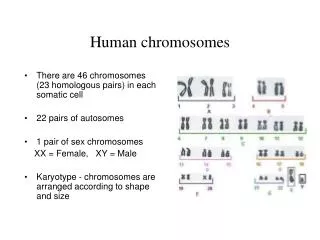
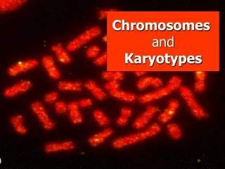
The Human Karyotype - Overview • Karyotypes show arranged chromosome pairs. • Standard karyotypes use the dye Giemsagenerating G-bands (gene poor regions vs R-bands). • Karyotypes are used in diagnosis. The chromosomes have been cut out of the photograph and paired with their homologs. The chromosomes as seen in the cell by microscopy.
Chromosome painting allows chromosome to display a unique color generating by a mix of many in situ hybridization probes. Chromosomes are then grouped in pairs and arranged in conventional order.
The five columns correspond to the five indicated fluorochromes and the 24 rows denote the 24 human chromosomes.
False colors representation of karyogram. Examples of arbitrary false colors: • Cy5 in Chromosome 1 is coded as white. • DEAC of Chromosome 2 coded as magenta. • TexRed of Chromosome 3 coded as light green. • FITC+SpeOra of Chromosome 12 coded as olive green and etc …. The Karyogram shows a pathologically serious defects due to human tumor (Adenom HRT 18): the chromosomes 8, 9, 14, 17, 22 are present only single. Chromosome 19 contains parts of 10 and 3 and Chromosome 22.
The short arm of a chromosome is called “p” and the long arm “q”. A through G refer to grouping of chromosomes by size.
Conventional symbols of chromosome abnormalities allows for ease of reference. Isochromosome 21 leads to Down Syndrome
The Human Karyotype – Centromere • Centromeres are crucial for cell division as they give stability to chromosomes. • Centromeres can be either localized or holocentric. • When localized, centromeres can lead to metacentric, submetacentricand acrocentric chromosomes. • A chromosome can lack centromere (acentric) or express two centromeres (dicentric).
The Human Karyotype – Dosage Compensation of X-linked Genes • In female mammals, one of two X chromosomes is inactivated in each cell. • Random process at about the 100-cell stage. • CondensedX is visible in normal cell as Barr body. • Normal females are mosaic(some cells XM, and some XP), where M = maternal chromosome, P = paternal.
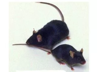
XIST (inactive X, Xi, specific transcripts) gene expressed in the X Inactivation Center (XIC) that generates a non-coding RNA. • Addition of methyl group on cytosine to CpG islands(DNA methyltransferase). • Histone code, enrichment of macroH2A. Calicocat, whose pattern is the result of random X inactivation. Alleles of X give orange or black (only one active X in each female cell); unlinked autosomal gene controls white spots.
RNA-fluorescence in situ hybridization detecting Xist RNA (red) localized on the inactive X in a preparation of condensed chromosomes from differentiated mouse cells
Trisomy X, Tetrasomy X and Pentasomy X Detection of the histone variant macroH2A in interphase nuclei from females with 46,XX, 47,XXX, 48,XXXX, and 49,XXXXX karyotypes. Regions of bright fluorescence indicate presence of macroH2A associated with inactive X chromosomes.
X-inactivation escape • Distribution along the human X chromosome of the approximately 15 percent of genes that escape complete transcriptional silencing in the inactive X chromosome. • Most are at PAR region(pseudo-autosomal) where X an Y pair at meiosis.
About 15% of genes escape X inactivation (random event, not same in all females), thus are expressed in both active and inactive X chromosomes. • Another 10% of genes may or may not escape X inactivation (Xp).
Chromosome Abnormalities in Human Pregnancies -Ploidyrefers to the number of sets of chromosomes in a cell. -Euploidyrefers to the state of a cell or organism having an integral multiple of the monoploid number, possibly excluding the sex-determining chromosomes. -Anaploidyrefers to not having euploidy.
Trisomy 21 (Down syndrome) is a viable condition, unlike most trisomies.
Trisomy 13, Patau syndrome, is a chromosomal condition associated with severe intellectual disability and physical abnormalities in many parts of the body. Trisomy 13 occurs in about 1 in 16,000 newborns microcephaly polydactyly
Trisomic Segregation In a trisomic organism, the segregation of chromosomes in meiosis is abnormal because the trisomic chromosome has two pairing partners. The trisomic chromosome can form a trivalentin some cells and one normal bivalent and one univalentin some other cells.
Chromosome Abnormalities in Human Pregnancies – Sex Chromosome Abnormalities Normal female (46, XX), Normal male (46, XY), Trisomy X (47, XXX), Double Y (47, XYY), Klinefelter syndrome (47, XXY) and Turner syndrome (45, X) (less viable).
If genetic recombination during meiosis happens outside of the pseudoautosomal region, any ensuing offspring will be either XX male or XY female. Metaphase chromosomes hybridized with LSI SRY probe
Chromosome Abnormalities in Human PregnanciesEnvironmental Effects on Nondisjunction Risk factors include radiation, alcohol, smoking, pesticides and other chemical such as bisphenol A.
Abnormalities of Chromosome Structure A, Terminal and interstitial deletions, each generating an acentric fragment. B, Unequal crossing over between segments of homologous chromosomes or between sister chromatids (duplicated or deleted segment indicated by the brackets). C, Ring chromosome with two acentric fragments. D, Generation of an isochromosome for the long arm of a chromosome. E, Robertsonian translocation between two acrocentric chromosomes. F, Insertion of a segment of one chromosome into a nonhomologous chromosome. Breakpoint can be mapped through testcrosses, thus generating genetic maps.
Chromosomal Deletion & Duplication - Deletions Deletions are a type of aberrations in chromosome structure and specifically refer to a chromosome having a missing segment of DNA. The severity of the phenotype depends on the missing length and the genotype of the individual. Deletions can arise from simple breakage and reunionor through ectopic recombinations.
Chromosomal Deletion & Duplication – Duplication Duplications are another type of aberrations in chromosome structure and specifically refer to a chromosome having a duplicated segments of DNA. Tandem duplication can have their own phenotypes (bar phenotype) Tandem duplication, through unequal crossing over, can produce more repeated sequences.
Red-green color blindness is and X-linked recessive disorder. • Unequal crossing over can occur in eye pigment genes (red-green) leading to color blindness. • Variation in the copy number of green visual pigment genes in the red and green visual pigment gene cluster on the X chromosome both in persons with normal color vision and in males with X-linked defects in green or red color perception. Organization of red-pigment and green-pigment genes in three wildtype X chromosomes.Origin of multiple green-pigment genes by unequal crossing over, lack of green-pigment gene in other chromosome.
Depending on the type of crossing over that occurs, several different types of chromosomal aberrations can occurs: • Lack of green pigment induces deuteranopia. • Chimeric genes induces deuteranopia or protomatopia.
Genetics of Chromosomal Inversions - Inversions Inversions are another type of aberrations in chromosome structure and specifically refer to a chromosome having a reversed order of genes in a particular DNA sequence. Inversions can occur through a two-break event or through ectopic recombination between inverted repeat sequences. During meiosis, heterozygous inversion leads to an inversion loop where the genes align at the prophase I synapsis complex.
If there is no crossing over within the inversion, then the homologous chromosomes separate normally during anaphase I.
Genetics of Chromosomal Inversions – Paracentric and Pericentric Inversions
Genetics of Chromosomal Inversions –Paracentric inversions If there is crossing over within the inversion and the inversion is paracentric, then the homologous chromosomes do not separate normally and dicentric and acentric chromosomes are generated.
Genetics of Chromosomal Inversions – Pericentric inversions If there is crossing over within the inversion and the inversion is pericentric, then the homologous chromosomes separate normally and monocentric chromosomes are generated, even if chromatids carry deletion and duplications of selected genes.
Chromosomal Translocations - Translocations Another frequently observed anomaly (1:1'000 newborns) is the robertsonian translocation, which occurs between two acrocentric chromosomes of groups G and D. It is also referred to as the centric fusion of two acrocentric chromosomes. It is a special kind of translocation in that on the acrocentric chromosomes (most often chromosomes 14 and 21 or 22) the very short, satellite-bearing arm is lost and a centric fusion t(14q21q or 14q22q) of the two remainder chromosomes, i.e., the long arms of the two pieces, results. In a reciprocal translocation two broken off chromosome pieces of non-homologous chromosomes are exchanged. This is a relatively frequent anomaly. One finds it with an incidence of 1:500 newborns. Reciprocal translocations are frequently balanced because the entire genetic material is present. Problems occur, though, in gamete formation.
Chromosomal Translocations - Reciprocal Translocations Meiosis in a translocation heterozygote has genetic complications called semisterility, as half the gametes are euploid (normal) and half of the gametes are aneuploid (abnormal) after the formation of the quadrivalent in the meiotic synapsis. Segregation of homologous chromosomes can occurs in three ways: Adjacent-1 or disjunctional (unbalanced gametes) Adjacent-2 or nondisjunctional (unbalanced gametes) Alternate (balanced gametes)
Chromosomal Translocations – Robertsonian Translocations A Robertsonian translocation can join chromosome 21 to any other acrocentric (ex. 14) chromosome; also causes Down syndrome. Heterozygous carriers of a Robertsonian translocation are phenotypically normal but a high risk of Down syndrome results from aberrant segregation in meiosis.
Genomic Position Effects on Gene Expression • Genes near breakpoints become repositioned and have new neighboring genes. • This repositioning may or may not affect the level of expression of these genes. • Position effect refers to an abnormal phenotype and position-effect variegation (PEV) is an example. Gene expression sometimes depends on the location of a gene in or near heterochromatin. Here patterns of red and white sectors in the eye of Drosophila melanogaster result from position-effect variegation.