Download
1 / 36
360 likes | 719 Views
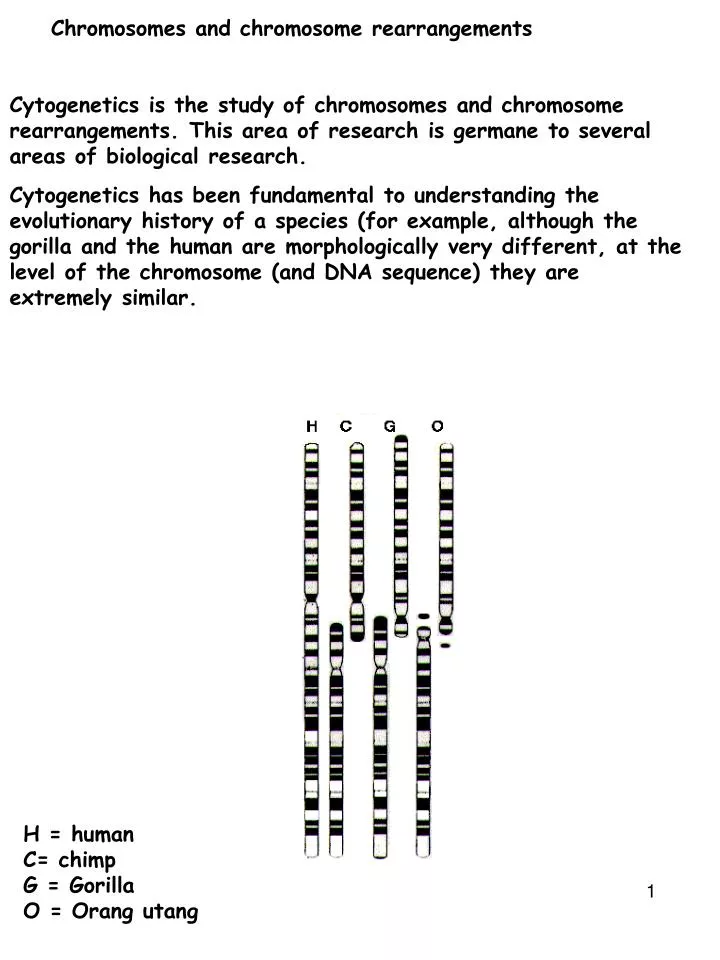
Chromosomes and chromosome rearrangements. Cytogenetics is the study of chromosomes and chromosome rearrangements. This area of research is germane to several areas of biological research.

E N D
Chromosomes and chromosome rearrangements Cytogenetics is the study of chromosomes and chromosome rearrangements. This area of research is germane to several areas of biological research. Cytogenetics has been fundamental to understanding the evolutionary history of a species (for example, although the gorilla and the human are morphologically very different, at the level of the chromosome (and DNA sequence) they are extremely similar. H = human C= chimp G = Gorilla O = Orang utang
Karyotype Chromosomes are classified by size, centromere position and banding pattern: Shown below is the human karyotype (description of the chromosome content of a given species) Karyotype is the chromosome description of length, number, morphology. Karyotype analysis is extremely important in medicine. Alternations in karyotypes are linked to birth defects and many human cancers. Metacentric- centromere in the middle Acrocentric- centromere off center telocentric centromere at one end
Banding patterns Specialized stains produce unique banding patterns along each chromosome. Banding patterns are extremely useful for detecting abnormalities in chromosome structure.
Gross chromosomal changes The Cri du chat syndrome in humans is a result of a deletion in the short arm of chromosome 5. This was determined by comparing banding patterns with normal and Cri du Chat individuals For many of the chromosome stains the molecular basis of the banding patterns is unclear. Nonetheless these techniques remain fundamental in many areas of genetic research Types of chromosome rearrangements that can be studied by karyotype analysis: GROSS CHROMOSOMAL CHANGES Deletions, Duplications, Inversions, Translocations
DDIT A____B____C________D____E____F Normal Chromosome Deletions (deficiency) Duplications Inversions Translocation
Deletions Deletions are often detected cytologically by comparing banding patterns between the normal and the partially deleted chromosomes Deleted chromosome Chromosome no female deletion chromosome1 Band 46,XX, del(1)(q24q31) Female with a deletion of chromosome 1 on the long arm (q) between bands q24 to q31.
In many instances deletions are too small to be detected cytologically. In these instances genetic criteria are used. Since deletions remove a contiguous set of genes, there is a high probability that an essential gene will be deleted. Therefore deletions will survive as heterozygotes and not homozygotes. Normal Homologous deletion (Lethal?) Heterologous deletion (NOT Lethal)
A+_____B+_____C+___________D+ Normal A+_____B+_____C+___________D+ In individuals heterozygous for the deletion, pairing is disrupted in the regions surrounding the deletion. Therefore recombination is also significantly reduced in these regions. Normal A deletion on one homologue unmasks recessive alleles on the other homologue. The effect is called pseudo-dominance. Hemizygous
Deletions in X Females in Drosophila XX Males in Drosophila XY or XO Deletion series phenotype
Changes in chromosome structure Deletions: Homozygosity for large deletions results in lethality- even the smallest cytologically defined deletions take out tens of 1,000's of bps and are likely to remove essential genes. 2. Organisms can tolerate heterozygosity for small but not large deletions. The reason for this is not entirely clear and is placed under the rubric of disrupting the overall ratio of gene products produced by the organism
Deletion mapping Deficiency mapping or deletion mapping: This provides a means of rapidly mapping a new mutation A deficiency or deletion is the loss of a contiguous series of nucleotides ATGATCGGGCCCATCAAAAAAAAAAAATCATCCCCCGGGG DELETION ATGATCGGGCCCATC CATCCCCCGGGG ATGATCGGGCCCATC|CATCCCCCGGGG Defined deficiencies are very useful for mapping genes
Deficiency mapping Generate a heterozygote Gene point mutant/deletion mutant Ask if you get intragenic recombinants Heterozygote will be pseudodominant The single point mutation will be observed over the deletion
Duplications A____B____C________D____E____F A____B____C________D____E____E____F normal Duplication Individuals bearing a duplication possess three copies of the genes included in that duplication. In general, for a given chromosomal region, organisms tolerate duplications much better than deletions. 46,XY, dup(7)(q11.2q22) Male with a duplication of chromosome 7 on the long arm (q) between bands 11.2 to 22.
Tandem duplications This is a case in which the duplicated segment lies adjacent to the original chromosomal segment A B C D ------ A B C B C B C D Once a tandem duplication arises in a population, even more copies may arise because of asymmetrical pairing at meiosis. Remember when the homologs pair during prophase of meiosis I, they line up base-pair for base pair. Duplications lead to mistakes in this pairing mechanism:
Proper pairing: A____B____C____B____C____D____E A____B____C____B____C____D____E A____B____C____B____C____D____E A____B____C____B____C____D____E Inappropriate pairing:
Proper pairing: A____B____C____B____C____D____E A____B____C____B____C____D____E A____B____C____B____C____D____E A____B____C____B____C____D____E Inappropriate pairing: A____B____C____B____C____D____E A____B____C____B____C____D____E A____B____C____B____C__-----------__D____E A____B____C____B____C__-----------__D____E
Tandem duplications expand by mistakes in meiosis during pairing
A B C B C D A B C B C D C B B C B A C B D A C D A B C B C D A B C B C B C D A B C D A B C B C D
The four meiotic products of a crossover between regions B and C: This process may repeat itself many times, such that a small fragment of the genome is repeated 10,000 times. An example of this is near the centromeres of the Drosophila genome: If you look at the DNA sequence in this region it consists of small 5-10 bp sequences (AATAC)n repeated 1,000s of times. It is believed to have arisen from unequal crossing over. Junk DNA Selfish DNA Conserved. Important?! Heterochromatin Genome sequence- Heterochromatin is usually not sequenced
Duplications provide additional genetic material capable of evolving new function. For example in the above situation if the duplication for the B and C genes becomes fixed in the population- the additional copies of B and C are free to evolve new or modified functions. This is one explanation for the origin of the tandemly repeated hemoglobin genes in humans. Each of these has a unique developmental expression pattern and provides a specialized function. The hemoglobin in fetus has a higher affinity for oxygen since it acquires its oxygen from maternal hemoglobin via competition
Some duplicated genes accumulate mutations and are no longer expressed (these are akin to junked cars along the highway). These are known as pseudogenes. One of the genes in the hemoglobin cluster is a pseudogene. -G-A-*-- pseudogene Unequal crossing over among the tandemly repeated hemoglobin gene cluster is the explanation for some inherited blood diseases. Hemoglobin lepore
Inversion Chromosomes in which two breaks occur and the resulting fragment is rotated 180 degrees and reinserted into the chromosome. Inversions involve no change in the amount of genetic material and therefore they are often genetically viable and show no abnormalities at the phenotypic level. Gene fusions may occur Inversions are defined as to whether they span the centromere Paracentric inversions do not span the centromere: Pericentric inversions span the centromere: In a pericentric inversion one break is in the short arm and one in the long arm. Therefore an example might read 46,XY,inv(3)(p23q27). A paracenteric inversion does not include the centromere and an example might be 46,XY,inv(1)(p12p31).
Homologs which are heterozygous for an inversion have difficulties pairing in meiosis. During pairing homologous regions associate with one another. Consequently individuals heterozygous for an inversion will form a structure known as an inversion loop. Crossover within inverted region? A---B---C---D---E---F---G A---B---C---D---E---F---G A’--B’---C’---D’--E’---F’--G’ A’--B’---C’--E’---D’--F’--G’
During meiosis, pairing leads to formation of an inversion loop This is a problem if crossing over occurs within the inversion As an exercise describe the consequence of crossover within a pericentric inversion (one that spans the centromere).
Paracentric inversion crosses over with a normal chromosome, the resulting chromosomes are an acentric, with no centromeres, and a dicentric, with 2 centromeres. • The acentric chromosome isn't attached to the spindle, so it gets lost during cell division, and the dicentric is usually pulled apart (broken) by the spindle pulling the two centromeres in opposite directions. These conditions are lethal. • Pericentric inversion crosses over with a normal chromosome, the resulting chromosomes are duplicated for some genes and deleted for other genes. (They do have 1 centromere apiece though). • The gametes resulting from these are aneuploid and do not survive. • Thus, either kind of inversion has lethal results when it crosses over with a normal chromosome. The only offspring that survive are those that didn't have a crossover. Thus when you count the offspring you only see the non-crossovers, so it appears that crossing over has been suppressed.
What are the consequences of crossing-over in an individual homozygous for an inversion? Genotype of an individual heterozygous for an inversion: Genotype of an individual homozygous for an inversion:
Translocations A segment from one chromosome is exchanged with a segment from another chromosome. Chromosome 1 A B C D E F ----------------------0----------------------- Chromosome 2 O P Q R S T ----------------------0----------------------- Reciprocal translocation A B C D S T ----------------------0----------------------- O P Q R E F ----------------------0----------------------- This is more specifically called a reciprocal translocation and like inversions (and unlike duplications and deficiencies) no genetic material is gained or lost in a reciprocal translocation.
long arms of chromosome 7 and 21 have broken off and switched places. So you can see a normal 7 and 21, and a translocated 7 and 21. This individual has all the material needed, just switched around (translocated), so they should have no health problems. However there can be a problem when this person has children. Remember that when the gametes are made, each parent gives one of each chromosome pair. What would happen if this person gave the normal seven and the 21p with 7q attached? t(11;18)(q21;q21) translocation between chromosomes 11 and 18 at bands q21 and q21 Philadelphia chromosome: t(9;22)(q34;q11).
long arms of chromosome 7 and 21 have broken off and switched places. So you can see a normal 7 and 21, and a translocated 7 and 21. This individual has all the material needed, just switched around (translocated), so they should have no health problems. However there can be a problem when this person has children. Remember that when the gametes are made, each parent gives one of each chromosome pair. What would happen if this person gave the normal seven and the 21p with 7q attached? There are three copies of 7q instead of two. And there is only one copy of 21q t(11;18)(q21;q21) translocation between chromosomes 11 and 18 at bands q21 and q21 Philadelphia chromosome: t(9;22)(q34;q11).
As with inversions, individuals heterozygous for a reciprocal translocation will exhibit abnormalities in chromosome pairing Notice this individual has the normal amount of genetic material (two copies of each gene). However it is rearranged. If the translocated fragment contains a centromere, you could get dicentri and acentric chromosomes How will translocated chromosomes pair in meiosis?
Homologous regions associate with one another. These chromosomes will follow Mendel's rule of independent of assortment. In this instance one must focus on the centromere There are two possible patterns of segregation.
Alternate segregation: • キN1 and N2 segregate to one pole • キT1 and T2 segregate the other pole • These gametes have the normal haploid gene content: one copy of each gene and are normal • Adjacent segregation: • キN1 and T1 segregate to one pole • キT2 and N2 segregate to the other pole • These gametes are anueploid: they are missing some genes and duplicated for other genes. • These forms of segregation are equally frequent. Therefore, in a translocation heterozygote, about 50% of the gametes are viable and 50% are unviable.
Reciprocal translocations result in genes that are known to map to different chromosomes but behave as linked genes. Under normal circumstances genes E and R assort independently because they are on different chromosomes. However in a translocation they will behave as closely linked genes and segregate together.
Translocations (and inversion) breakpoints sometimes disrupt an essential gene. That is the break occurs in the middle of a gene. In fact because of this, a number of specific translocations are causally associated with specific human cancers. The inherited disease Duchenne muscular dystrophy was mapped through a translocation that specifically disrupted this gene.
abl/bcr Fusion protein Chronic myelogenous and acute lymphotic leukemia ALK/NPM Fusion Large cell lymphomas bcr/abl Fusion Chronic myelogenous/acute lymphotic leukemia HER2/neu Fusion Breast and cervical carcinomas MYH11/CBFB Fusion Acute myeloid leukemia ML/RAR Fusion Acute premyelocytic leukemia ERG/TMPRSS2 Fusion prostate cancer Gene fusion -prostate cancer -ERG merges with a prostate-specific gene called TMPRSS2. ERG is a transcription factors