Download

1 / 24
250 likes | 442 Views
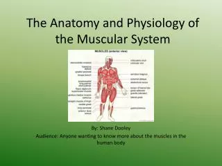
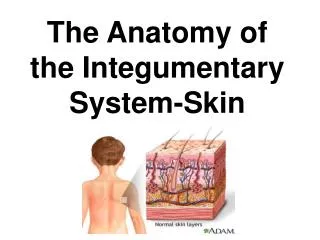
ANATOMY AND PHYSIOLOGY OF THE INTEGUMENTARY SYSTEM. OBJECTIVES. DEFINE INTEGUMENTARY SYSTEM AND IDENTIFY THE FUNCTIONS OF SKIN LEARN THE STRUCTURE AND FUNCTIONS OF THE SKIN’S ACCESSORY ORGANS RECOGNIZE AND DIFERENTIATE BETWEEN THE VARIOUS SKIN LESIONS LEARN THE EFFECTS OF UV LIGHT ON THE SKIN

E N D
OBJECTIVES • DEFINE INTEGUMENTARY SYSTEM AND IDENTIFY THE FUNCTIONS OF SKIN • LEARN THE STRUCTURE AND FUNCTIONS OF THE SKIN’S ACCESSORY ORGANS • RECOGNIZE AND DIFERENTIATE BETWEEN THE VARIOUS SKIN LESIONS • LEARN THE EFFECTS OF UV LIGHT ON THE SKIN • LEARN SOME COMMON DISEASES OF THE SKIN AND SKIN’S ACCESSORY ORGANS
INTRODUCTION • Skin is classified as both a body organ and a body system • An adult’s skin covers 3000 sq. inches • Makes up 15% of body weight • Destruction of 1/3 or more may result in death • Forms a continuous covering of a body
FUNCTIONS OF SKIN • PROTECTION • When intact protect against bacteria, sun, lass of fluids • TEMPERATURE CONTROL • Sweat glands cool down the skin • Dilation and constriction of blood vessels • PERCEPTION • Receptors for pain, touch, pressure are located in the skin • STORAGE, EXCRETION, SYNTHESIS • Stores fat • Synthesis of vit D • Excretes salts, water, some waste products
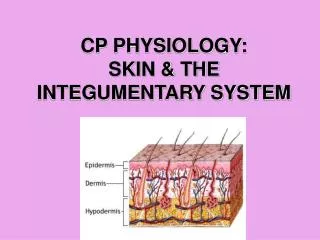
STRUCTURE OF THE SKIN • EPIDERMIS • DERMIS • SUBCUTANEOUS TISSUE
EPIDERMIS The top layer of skin Surface cells constantly flake off and are completely replaced every 30-45 days New cells are formed deep in the epidermis Skin color is determined by pigment melanin Melanocytes are cells producing melanin to protect against UV light.
SKIN DISCOLORATION • Erythema – red discoloration of skin • Cyanosis- bluish discoloration due to lack of oxygen • Jaundiceis a yellow color of the skin, mucus membranes, or eyes. The yellow coloring comes from bilirubin • Yellow skin due to too much caroten • Pallor – very pale skin • Melasma (also known as chloasma) is characterized by tan or brown patches that may involve the forehead, cheeks, upper lip, nose, and chin. Although this condition is typically termed the "pregnancy mask," men can also develop it
DERMIS Dermis is middle layer of skin Contains blood vessels, nerves, hair follicles, glands Dermis consists of connective tissue fibers – elastin, collagen
HYPODERMIS OR SUBCUTANEOUS TISSUE • The subcutaneous tissue is the third of the three layers of skin. • The subcutaneous layer contains fat and connective tissue that houses larger blood vessels and nerves The hypodermis is used mainly for fat storage.
ACCESSORY SRTUCTURES OF THE SKIN • HAIR • SWEAT GLANDS • SEBACEOUS GLANDS • NAILS
HAIR • Hair is composed of strong structural protein called keratin. • Below the surface of the skin is the hair root, which is enclosed within a hair follicle. • The hair follicle contains pigment cells that produce melanin. Melanin is the chemical that gives hair its color. • White hair is caused by air rather than pigment in melanocytes • Scalp hair grows at average rate of 1mm every 3days.
SWEAT/SUDORIFEROUS GLANDS • There are between 2 and 4 million sweat glands found across the human body. • These are coiled tubular glands that are found in the dermis or lower part of the skin. They produce a watery secretion and open on to the skin to help control body temperature . • When you sweat the moisture on your body evaporates and provides a cooling effect for the body.
SEBACEOUS GLANDS • The sebaceous glands are microscopic glands in the skin that secrete an oily/waxy matter, called sebum, to lubricate and waterproof the skin and hair. • They are distributed throughout all skin sites except the palms and soles. • Sebaceous glands secrete the oily, waxy substance called sebumthat is made of fat, wax, and the debris of dead fat-producing cells.
NAILS • The fingernail is an important structure made of keratin that has 2 purposes. • The fingernail acts as a protective plate • Enhances sensation of the fingertip. • The structure we know of as the nail is divided into six specific parts: • the root • nail bed • nail plate • eponychium(cuticle) • perionychium • hyponychium
INFLAMMATORY DISEASES • Acne Vulgaris • Acne is a skin condition that causes whiteheads, blackheads, and inflamed red growths (papules, pustules, and cysts) to form • Acne occurs when tiny holes on the surface of the skin, called pores, become clogged. • Each pore is an opening to a follicle, which contains a hair and an oil gland. These oil glands help lubricate the skin and help remove old skin cells. • When glands produce too much oil, the pores can become blocked. Dirt, debris, bacteria, and inflammatory cells build up. The blockage is called a plug or comedone • The top of the plug may be white whitehead or dark blackhead.
Impetigo – Very contagious, superficial skin infection usually seen in young children. • Cause – bacterial infection – streptococcus or staphycoccocus • Symptoms – small red macule turns into a vesicle, then a pustule. Lesions break and yellow crust forms. • Treatment – good hygiene, topical antibiotics • Folliculitis – Bacterial infection of the hair follicles • Furuncles – hard, painful nodules that enlarge and rupture
Herpes • Herpes Simplex I – cold sores • Vesicles or blisters of the mouth, nose, skin • Treatment – topical application of antiinflammatorydrugs • Herpes Zoster – same as chicken pox • Fever, severe pain in affected areas, vesicalerruptions • Treatment – analgesics, antipyretics, immunostimmulators
SKIN CANCER • Classification: • Basal cell carcinoma • Squamous cell carcinoma • Melanoma • Basal cell carcinoma is the most common form of skin cancer and accounts for more than 90% of all skin cancer in the U.S. • These cancers almost never spread (metastasize) to other parts of the body. • Risk factors for developing basal cell carcinoma
age • exposure to ultraviolet radiation • exposure to sun • therapeutic radiation • Malignant Melanoma is the most aggressive of malignant cutaneous tumors. • Cases with lymphnodeinvolvement, and distant metastases, carry a very poor prognosis, (50% and 20% respectively alive in 5 years),
TREATMENT • Four types of standard treatment are used: • Surgery • Radiation therapy • Chemotherapy • Photodynamic therapy • New types of treatment are being tested in clinical trials. • Biologic therapy
ABCDs of Melanoma • A = Asymmetry: Moles are commonly symmetrical and round. Very early melanomas, are asymmetrical assuming different forms. One half does not match the other half.B = Border: Commonly moles, have even and smooth borders. Early melanomas, are usually uneven and notched borders.The edges are ragged, notched or blurred.C = Color: Moles commonly are a single shade of brown, early malignant melanomas shows brown-tan or black color. Further, as melanomas progress, may see red, white, and blue color.D = Diameter: Early malignant melanomas, differently from moles, tend to grow larger in diameter. First suspects in melanoma start from a 6 millimeters diameter on. Any sudden or continuing increase in size should be of special concern.