Download
1 / 20
270 likes | 901 Views
Urinary System- Anatomy and Physiology. Zoe McCarthy. Urinary System in Context. Urinary System in Context. Functions of the Urinary system. 1. Regulating blood volume and pressure 2. Regulating plasma concentrations of sodium, potassium, chloride and other ions 3. Stabilising blood pH

E N D
Urinary System-Anatomy and Physiology Zoe McCarthy
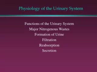
Functions of the Urinary system • 1. Regulating blood volume and pressure • 2. Regulating plasma concentrations of sodium, potassium, chloride and other ions • 3. Stabilising blood pH • 4. Conserving nutrients • 5. Detoxifying poisons (with the liver)
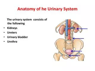
Organisation of the Urinary System • Kidneys • Ureters • Urinary bladder • Urethra
Position of the Kidneys CT abdomen with contrast MRI coronal abdomen
Protection of the Kidneys • 3 layers of connective tissue: • Inner layer- Renal capsule • Middle layer- Adipose capsule • Outer layer-Renal fascia Renal cortex Retroperitoneal space
Surface anatomy of the Kidney • Hilum is located on the medial surface 10 cm 3cm 5.5cm
Internal Structure of the Kidney Renal Lobe Renal pyramids Renal papilla Renal Columns
Nephron-Tubular System • Proximal convoluted tubule • Descending loop of Henle • Ascending loop of Henle • Distal convoluted tubule • Collecting duct
Summary so far….. Blood enters the kidney through the renal artery at the site of the hilum The renal artery divides in to ever smaller arteries and arterioles Afferent arterioles take blood to the glomerulus to be filtered Once blood is filtered efferent arterioles take blood away from the glomerulus The PCT is concerned with reabsorption- organic nutrients are reabsorbed and water follows because there is a concentration gradient The filtered substances move into the proximal convoluted tubule Products which are filtered out: water, mineral salts, amino acids, glucose, hormones, urea, toxins The glomerulus is a network of capillaries which filters the blood Products which do not filter and remain in the blood: Leukocytes, erythrocytes, platelets, plasma proteins The remaining filtrate moves into the descending loop of henle. This is lined with thin cells so water moves out Because water has been reabsorbed the concentration of the filtrate is not very high The walls of the ascending loop of henle are lined with thicker cells, so water can’t pass in or out. Instead sodium and chloride is pumped out actively The filtrate now enters the distal convoluted tubule- is it now only 20% of what it originally was. A number of other nephrons join up to the cleectig duct which travels through the medulla to the renal papilla wher the filtrate is emptied in the minor calyx In the DCT the volume and composition of the filtrate can be adjusted but this is controlled by hormones From the DCT the filtrate now passes into the collecting duct. 4-5 minor calyces join up to make a major calyx 2-3 major calyces join up to form the renal pelvis The renal pelvis joins the ureter at the hilum The ureter transport the filtrate/urine from the kidney to the bladder
The Formation of Urine • 3 processes involved in the formation of urine. • Simple filtration • Selective reabsorbtion • Hormonal control- • Parathyroid hormone, calcitonin • Anti diuretic hormone • Aldosterone • Secretion
Ureters • Superiorly • Continuous with the renal pelvis • Inferiorly • Pass through the abdominal cavity, behind the peritoneum, infront of the psoas muscle, into the pelvic cavity ehere they enter the posterior wall of the bladder • 25-30 cm in length
Ureter- Cross Section • 3 layers of tissue • Outer layer • Fibrous tissue • Middle layer • Muscle • Inner layer • Epithelium
Bladder- structure of • 3 layers • Outer layer • Loose connective tissue • Middle layer • Smooth muscle and elastic fibres • Inner layer • Lined with transitional epithelium
Urethra • Extends from the base of the bladder to the outside world. • Anatomical differences mean that male and female urethras are different. • Female: 4cm long • Male: 14cm long
Urethra- structure of • Muscle layer • Submucosa layer • Mucosa