Download
1 / 126
1.33k likes | 1.69k Views
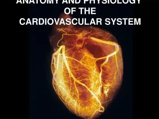
Anatomy and Physiology of the Cardiovascular System. Prepared by Miss Fatima Hirzallah. The heart is a hollow, muscular organ situated in the space between lungs(mediastinum) , its about 12 cm in length & about 9 cm in width. Cardiac Muscle . Contract as a single unit

E N D
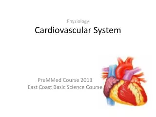
Anatomy and Physiology of the Cardiovascular System Prepared by Miss Fatima Hirzallah
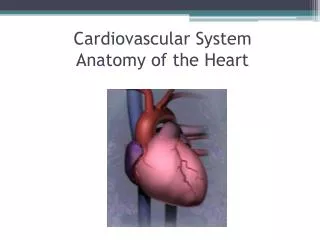
The heart is a hollow, muscular organ situated in the space between lungs(mediastinum) , its about 12 cm in length & about 9 cm in width
Cardiac Muscle • Contract as a single unit • Simultaneous contraction due to depolarizing at the same time • Automaticity
The heart is about the size of a clenched fist and comprises. • The heart composd of four layers: • Endocardium, • Myocardium, • Epicardium, • and the pericardium..
endocardium is the inner layer and is consists of endothelial tissue that lines the inner surface of the heart and the cardiac valves. • The myocardiumis the middle layer and is composed of muscle fibers that enable the heart to pump. • Epicardium is the outer layer, is tightly adherent to the heart and the base of the great vessels. • A thin, fibrous, double-layered sac known as the pericardiumsurrounds the heart.
The outer layer is known as the parietal pericardium • and the inner layer is called the visceral pericardium • Between these two layers is a small amount of pericardial fluid (30 to 50 mL) that serves as a lubricant between the two layers
The heart consists of four chambers: • right and left atrium • right and left ventricles.
Heart valves • The cardiac valves are composed of fibrous tissue and allow blood to flow in one direction. • The valves open and close as a result of blood flow and pressure differences.
The tricuspid and mitral valves are known as the atrioventricular (AV) valves because they are located between the atria and the ventricles. • The pulmonic and aortic valves are known as the semilunar valvesbecause each has three leaflets shaped like half-moons.
Circulation of the blood • The blood passes through the tricuspid valve into the right ventricle, which then pumps the blood through the pulmonic valve into the pulmonary circulation. • After gas exchange in the lungs, oxygenated blood returns to the left atrium, passes through the mitral valve, enters the left ventricle, passes through the aortic valve, and finally enters the aorta
Coronary Arteries • The left and right coronary arteries and their branches supply arterial blood to the heart. These arteries originate from the aorta just above the aortic valve leaflets. • The heart has large metabolic requirements, extracting approximately 70% to 80% of the oxygen delivered (other organs consume, on average, 25%).
The left coronary artery has three branches. 1-the artery from the point of origin to the first major branch is called the left main coronary artery. two bifurcations arise off the left main coronary artery 2- left anterior descending artery (LAD), which courses down the anterior wall of the heart 3-circumflex artery, which circles around to the lateral left wall of the heart.
The right side of the heart is supplied by the right coronary artery, which progresses around to the bottom or inferior wall of the heart. • The posterior wall of the heart receives its blood supply by an additional branch from the right coronary artery called the posterior descending artery.
The coronary arteries are perfused during diastole. An increase in heart rate shortens diastole and can decrease myocardial perfusion. • Patients, particularly those with coronary artery disease (CAD), can develop myocardial ischemia (inadequate oxygen supply) when the heart rate accelerates.
Cardiac Output • Cardiac output is the amount of blood pumped out of the ventricle . • The cardiac output in a resting adult is about 5 L per minute but varies greatly depending on the metabolic needs of the body. Cardiac output is computed by multiplying the stroke volume by the heart rate.
Stroke volume (SV) :The amount of blood ejected by the left ventricle with each heartbeat . • the heart rate is 60 to 80 beats per minute (bpm) • The average resting stroke volume is about 70 mL, and Cardiac output can be affected by changes in either stroke volume or heart rate.
Cardiac Output/Index • Cardiac output • CO = HR (beats/minute) X SV (liters/beat) • Normal adult: 4-8 liters/minute • Cardiac index • CI = CO(liter/minute)/Body surface area (m2) • Normal adult: 2.8-4.2 liter/minute/m2 • Normalizes liter flow to body size
Stroke Volume • Preload • Afterload • Contractility
Stroke Volume • Preload • The amount of stretch placed on the cardiac muscle just prior to systole (the amount of the ventricle at end diastole) • Diastole : filling stage of cardiac cycle. • Afterload • The force or pressure at which the blood is ejected from the left ventricle • Equated with systemic vascular resistance (SVR)
Contractilityis a term used to denote the force generated by the contracting myocardium under any given condition • The resistance of the systemic BP to left ventricular ejection is called systemic vascular resistance. • The resistance of the pulmonary BP to right ventricular ejection is called pulmonary vascular resistance .
The percentage of the end-diastolic volume that is ejected with each stroke is called the ejection fraction (EF) (EF) = 50-70%
HEALTH HISTORY AND • CLINICAL MANIFESTATIONS For the patient experiencing an acute MI, the nurse obtains the health history using a few specific questions about the onset and severity of chest discomfort, associated symptoms, current medications, and allergies. At the same time, the nurse observes the patient’s general appearance and evaluates hemodynamic status (heart rate and rhythm, BP).
Cardiac Signs and Symptoms • Chest pain or discomfort (angina pectoris, MI, valvular heart disease) Shortness of breath or dyspnea (MI, left ventricular failure, HF) • Edema and weight gain (right ventricular failure, HF) • Palpitations (dysrhythmias resulting from myocardial ischemia, stress, electrolyte imbalance)
• Fatigue (earliest symptom associated with several cardiovascularndisorders) • Dizziness and syncope or loss of consciousness (postural hypotension, dysrhythmias, vasovagal effect,cerebrovascular disorders)
Inspection General appearance Jugular venous distension (JVD) Skin Extremities Palpation Pulses Point of maximal impulse (PMI) Percussion Auscultation Good stethoscope Positioning Normal tones – S1/S2 Extra tones – S3/S4 Murmurs Rubs Physical Exam
HEART SOUNDS HEART SOUNDS The normal heart sounds, S1 and S2, are produced primarily by the closing of the heart valves. The time between S1 and S2 corresponds to systole This is normally shorter than the time between S2 and S1 (diastole). As the heart rate increases diastole shortens. S1—First Heart Sound. Closure of the mitral and tricuspid valves creates the first heart sound (S1), S2—Second Heart Sound. Closing of the aortic and pulmonic valves produces the second heart sound (S2).
Murmurs are created by the turbulent flow of blood. • The causes of the turbulence may be a critically narrowed valve, • a malfunctioning valve that allows regurgitant blood flow, • a congenital defect of the ventricular wall, a defect between the aorta and the pulmonary artery,
Diagnostic Evaluation • Laboratory test(Cardiac Labs) • Chest X-ray • ECG • CARDIAC STRESS TESTING • ECHOCARDIOGRAPHY(ECO) • Echocardiography is a noninvasive ultrasound test that is used to examine the size, shape, and motion of cardiac structures.
Important Cardiac Labs • Enzymes – CK, CK-MB, LDH • Other important cardiac biomarkers that are assessed include the myoglobin and troponin T or I. Myoglobin
early marker of MI, is a heme protein with a small molecular weight. This allows it to be rapidly released from damaged myocardial tissue and accounts for its early increase, within 1 to 3 hours after the onset of an acute MI. Myoglobin peaks in 4 to 12 hours and returns to normal in 24 hours.
Lipid studies – Cholesterol, triglycerides Coagulation studies – PTT and PT/INRI (nternational • Normalized Ratio (INR).The INR provides a standard method for reporting PT level • Electrolytes – Potassium, magnesium, and calcium
Invasive Tests • Cardiac catheterization • Coronary angiography
To pump effectively, large portions of cardiac muscle must receive an action potential nearly simultaneously. • Special cells that conduct action potentials extremely rapidly are arranged in pathways through the heart.
Before mechanical contraction, an action potential travels quickly over each cell membrane and down into each cell’s.
Three physiologic characteristics of two specialized electrical cells, the nodal cells and the Purkinje cells, provide this synchronization: • Automaticity: ability to initiate an electrical impulse • Excitability: ability to respond to an electrical impulse • Conductivity: ability to transmit an electrical impulse from one cell to another
Cardiac Conduction • Sinoatrial (SA) node – Fires at 60–100 beats/minute • Intranodal pathway • Atrioventricular (AV) node – Fires at 40-60 beats/minute • Atrioventricular bundle of His • Ventricular tissue fires at 20-40 beats/minute and can occur at this point and down • Right and left bundle branches • Purkinje fibers
12-Lead ECG • Limb leads • Standard leads: I, II, and III • Augmented leads: aVR, aVL, and aVF • Precordial leads • V1,V2,V3,V4,V5, and V6 • Axis • The direction of the flow of electricity