Download
1 / 44
470 likes | 1.3k Views
Chapter 3 Digital Radiography . Outline. Medical Imaging Modalities (X-ray, CT, MR, US,; Computed Radiology, Digital Radiology) Image Quality (S/N, Spatial Resolution, Density Resolution, Window Adjust) Image Compression. /. /. Modalities Evolution. //. vacuum. semiconductors. networks.

E N D
Outline • Medical Imaging Modalities (X-ray, CT, MR, US,; Computed Radiology, Digital Radiology) • Image Quality (S/N, Spatial Resolution, Density Resolution, Window Adjust) • Image Compression
/ / Modalities Evolution // vacuum semiconductors networks tubes discrete components integrated circuits digital thermonic tubes X-ray image intensifier Digital radiography Rontgen Radiograph Casson ECT SPECT PET Nuclear Medicine pulsed Doppler Hertz Soldner CFI real time Ultrasound Hounsfield CT Lauterbur MRI Source: Brueckner E.K., Siemens Medical Systems Erlangen
Classification of Medical Image • 侵入性檢查 • 心導管 • 內視鏡 • 非侵入性檢查 • X光 (Radiograph) • 電腦斷層 (CT) • 磁振造影 (MRI) • 動脈攝影 (DSA) • 超音波 (US) • 核醫造影 (NM) • 眼底螢光攝影 (FA)
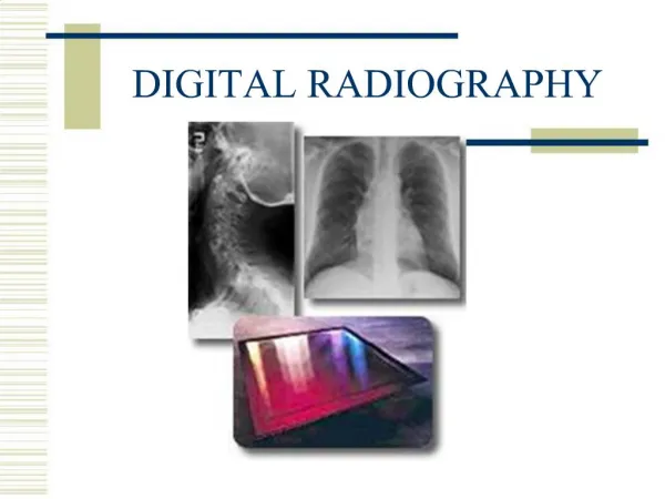
Radiology Technique of Digital Medical Image • 投影式造影(Projection Radiography) • X光片掃描機 (X-film Scanner) • CR (Computed Radiography) • DR (Direct Radiography) • DF (Digital Fluorography) • 斷層影像重組 • CT (Computed Tomography, 電腦斷層) • MR (Magnetic Resonance, 核磁造影) • Ultrasound (超音波) • 其他:Nuclear Medicine, Microscopic Imaging
X-ray Procedure ROOM X-ray tube Patient table 2 12 5 6 X-ray control console Bucky tray X-ray Generator 4 Diagnostic Area 9 10 11 Film viewing and Diagnosis area (light box) Un- Exposed Film screen Cassette storage 3 7 8 1 13 Film processing Area (dark room) X-ray Requisition Dispatcher and patient Escort Area
Plastic Bundle HE-NE Laser Tube Photo Sensor Mirror Beam Expander Cylindrical Lens Film Polygonal Mirror (polygon) F- lens Mirror X光片掃描機 (X-film Scanner)
雷射光電子掃描裝置 Mechanical Controller HOST computer CPU Parallel Computer Interface He-NE la3er Data Controller FLM 12BIT Log Amplifier A/D converter P.M.T Rotating polygonal Mirror Optical FLBER Light COOPLING Bundle X光片掃描機 (X-film Scanner)
A/Dconverter Semiconductor laser controller The Host computer CRT Stimulable phosphor detetor Schematic showing formation of the latent image on the CR imaging plate (1) the plate being scanned by the laser beam (2) light photons being converted to electrical signal (3) and electrical signals being converted to digital signals, which form a CR image (4) Conrtesy of Konica Corporation) CR (Computed Radiography)
Unused Imaging Plate BaFX Crystals Support X-ray photons X-ray Eeposure Recording the X-ray Imaging Laser-Beam Scanning The Laser Beams Extracts the X-ray Image from the plate by Converting to Light Photo, which from a Light Image Reading Light The Small Amount of Residual Image on the Plate is Thoroughly Erased by Flooding the Plate with Light Erasing The Erase Plate Can Be Used Again CR (Computed Radiography)呈像原理
(1) (2) (3) (4) TVMemter Image Intensifier Tube TV Camera Digital Chain Optics Grid X-ray table Table Patient Image Intensifier Tube Collimator DF (Digital Fluorography)
Projection data Direction of projection Cross section of the object 0 s Y θ 0 X Cross section of an imaged object, and projected in θ direction. ƒθ (S) shows projection data CT (Computed Tomography)電腦斷層呈像原理
1 1 5 5 10 10 15 15 20 20 25 25 30 30 Gantry Rotation 30sec Pitch=1:1 with 1cm collimation the bed will advance 30cm in 30sec (A) SINGLE HELICAL MODE Gantry Rotation 15sec 7sec NO Movement 15sec PATIENTS BED MOVEMENT PATIENTS BED MOVEMENT PATIENTS BED MOVEMENT (B) CLUSTER HELICAL MODE Helical(spiral) CT scanning modes. CT (Computed Tomography)
Display System Video monitor Image processor D/A Display system matrix camera Tape Drive D/A Digital Domain Computer Disk storage ControlInterface A/D Signal Demodulator RF Transmitter X-gradient Power Supply Y-gradient Power Supply Z-gradient Power Supply RF Receiver RF Power Amp Pre-amp Magnet Transmitting / Receiving Coil Y Magnet X-gradient coil Y-gradient coil Z-gradient coil X Z MR (Magnetic Resonance)核磁造影系統架構
Transmit Clock Pulse (PRF) Digital Scan Converter TGC Circuit HV Pulse Generator Transmitter Circuit Receiver Circuit Video Display Position Encoder Circuit Transtucer Ultrasound (超音波)系統架構
Origin y f(x,y) x 數位影像表示法 • 像素 (Pixel) • 解析度(Resolution) • Spatial Resolution • row x column • 灰階值(Gray level) • Density Resolution • 彩色:R(ed), G(reen), B(lue) • 影像大小(image size) • row x column x bits(實務上,影像多半以bytes為儲存單位;例如,12bit 影像就以 2byte 儲存)
各式醫學影像之大小 Row X Col X bits # of images/ Exams M byte/ Exams Modality Nuclear Medical (NM) Magnetic Resonance Imaging (MRI) Ultrasound (US) Digital subt. Angiography (DSA) Digitized Electronic Microscopy Digitized Color Microscopy Computed Tomography (CT) Computed Radiography (CR) Direct Radiography (DR) Digitized X-Rays Digital Mammography 128 X 128 X 12 256 X 256 X 12 512 X 512 X 8(24) 512 X 512 X 8 512 X 512 X 8 512 X 512 X 24 512 X 512 X 12 2048 X 2048 X 12 2048 X 2048 X 12 2048 X 2048 X 12 4000 X 5000 X 12 30-60 60 20-230 15-40 1 1 40 2 2 2 4 1-2 8 5-60 4-10 0.26 0.79 20 16 16 16 160
色彩基本原理(一) 加色原理 (螢幕) 減色原理 (印表機) 基本色:Red、Green、Blue 基本色:Red、Blue、Yellow
色彩基本原理(二) 一般電腦螢幕能顯示最多256層灰階變化 ( R , G , B ) ( 255, 0, 0) ( 0, 255, 0) ( 0, 0, 255) ( 0, 0, 191) ( 255, 255, 0) ( 191, 191, 0) ( 127, 127, 0) ( 63, 63, 0) ( 0, 0, 0) ( R , G , B ) ( 255, 255, 255) ( 223, 223, 223) ( 191, 191, 191) ( 159, 159, 159) ( 127, 127, 127) ( 95, 95, 95) ( 63, 63, 63) ( 31, 31, 31) ( 0, 0, 0) 灰階變化
影響顯像品質之要素 • 空間解析度(Spatial Resolution) • 影像大小(由儀器設備端決定) • 影像壓縮:Lossy or loseless • 畫質解析度(Density Resolution) • 灰階值的範圍(由儀器設備端決定) • 影像明暗對比:用Window level / width調整 • 訊雜比(Signal-to-Noise Ratio)
影像放大 (Zoom In) 原圖直接放大會有 Aliasing 現象發生(即顯現大顆粒格子狀的影像) 放大 8 倍 原影像 放大 64 倍
影像平滑處理 (Smoothness) Anti-Aliasing 未平滑處理 經平滑處理
影像平滑處理(二) 心得:影像中看到的邊緣(Edge)就是灰階突然變化之處, 平滑處理就是將灰階的變化變得較為平緩。 灰階值 灰階值 利用內插法(Interpolation)來處理 未平滑處理 平滑處理
局部處理 大範圍處理 影像平滑處理(三) 未平滑處理 平滑處理(一) 平滑處理(二)
Brightness and Contrast • Histogram: • # of pixels vs pixel value • Window level (Center) • Level 值變大→ 亮度變暗 • Level 值變小→ 亮度變亮 • Window width • width 值變大→ 對比變弱 • width 值變小→ 對比變強
影像濾波處理(Filter): (1) 原影像 Spatial Domain Frequency Domain
影像濾波處理(Filter): (2) 低頻譜影像 Spatial Domain Frequency Domain
影像濾波處理(Filter): (3) 高頻譜影像 Spatial Domain Frequency Domain
Image Quality Control • How to show images in the best quality without user’s manipulation ? • Image parameters (e.g.window width/level) of automatically-sent images may be not optimized by technologists. • cp. Frame-grab images are always optimized. • Using quality-control workstations to audit the image quality.
Specifications of the monitor • 0.25 mm focal point • Resolution 1024x768 for VGA non-interlaced • Max. refresh rate: 89 Hz • H.F.: 68 kHz V.F.:85 Hz
Quality Control Methods • Preset a window table on the PACS server • window width and level value. • Retrived image and table matching. • a field of image header . • Window re-computing during transmission.
CT SPIRAL_THORAX=450,40 CHEST_GENERAL=432,8 ADULT_BRAIN=400,50 CHILD_BRAIN=230,60 SPIRAL_BRAIN=400,50 SPIRAL_ABDOMEN=300,40 MEDIUM_abdomen=400,60 CR CHEST_PORTABLE=570,380 CHEST_GENERAL=720,470 ABDOMEN_GENERAL=630,550 CHEST_A_P=480,440 PELVIS_GENERAL=550,550 FOOT=650,350 IVP:C=640,590 Display parameters table
Goal of Image Quality Assuring • Optimal images were displayed on viewing station. • Quality was improved on every image modalities. • Acceptable by clinical users and radiologists. • Image contrast higher than the original image (narrower dynamic range) . • Automatic and expeditious image re-computing after window width and level matching.
Supplements to the display of image windows • Automatic adjustment • Presetting value (Icon) for bone, lung and soft tissue (CT). • Manual adjustment • Adjustable window width and level on histogram.
Image Compression 影像壓縮法 非失真型壓縮法: 不允許解壓資料產生誤差 文字壓縮 LZW, ZIP, ARJ 壓縮比約為2至5倍 失真型壓縮法: 解壓資料可以不全等於原始資料 video, still image, music JPEG, MPEG, H.263, H.324 壓縮比約為8至20倍
ROI Compression (a) Original Image (b) ROI Block (c) After Compression
Image Compression • 已將JPEG 2000定為DICOM標準 • 分為8/10/12/16/32 bit的JPEG lossy /lossless壓縮格式 • DICOM JPEG 格式為 • 70(即 JPEGlossless default ) • 50與51(即JPEG lossy default) • Lossy Compression will cause untrusty PACS