Download
1 / 33
750 likes | 1.27k Views
Peritonitis. Intra-abdominal infections Two major clinical manifestations Early or diffuse infection results in localised or generalised peritonitis Bacterial peritonitis is classified as primary or secondary Late and localised infections produces an intra-abdominal abscess

E N D
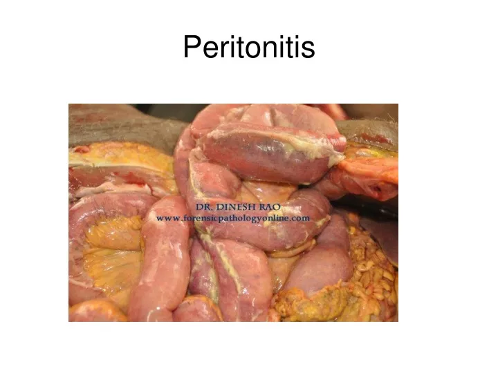
Intra-abdominal infections Two major clinical manifestations Early or diffuse infection results in localised or generalised peritonitis Bacterial peritonitis is classified as primary or secondary Late and localised infections produces an intra-abdominal abscess Pathophysiology depend on competing factors of bacterial virulence and host defences Peritonitis
Primary peritonitis • Diffuse bacterial infection without loss of integrity of GI tract • Often occurs in adolescent girls • Streptococcus pneumonia commonest organism involved • Ascites, secondary to cirrhosis of the liver, may become infected spontaneously. • Secondary peritonitis • Acute peritoneal infection • Often involves multiple organisms - both aerobes and anaerobes • Commonest organisms are E. coli and Bacteroides fragilis
Etiology (secondary peritonitis ) • Perforation of a viscus into the peritoneal cavity • Trauma • Infected intraperitoneal blood from any source (eg, trauma, surgery, ectopic pregnancy) can become infected and lead to peritonitis. • Foreign bodies • Pancreatitis
Etiology (secondary peritonitis ) • Strangulatingintestinal obstruction • Pelvic inflammatory disease (PID) • Vascular catastrophes (mesenteric thrombosis or embolism). • In sexually active women: gonococcus and chlamydia are most common. • IUD long-lasting • Anastomotic dehiscence
Etiology (secondary peritonitis ) • Peritoneo-systemic shunts, in common with other long-lasting • peritoneal drains, tend to become infected and lead to peritonitis. • Drains of any type may furnish an entry for bacteria into the peritoneal cavity. • Barium introduced into the peritoneal cavity via an enema through a perforated diverticulum can lead to acute and later to chronic peritonitis because of the combination of barium and infection. • Meconium peritonitis can occur from perforation of the bowel in utero. • Peritoneal dialysis
Peritonitis, symptoms • Abdominal pain • Abdominal tenderness • Fluid in the abdomen • Inability to pass feces or gas („ silent „ abdomen ) • Distended abdomen • Fever • Low urine output • Nausea and vomiting • Point tenderness • Thirst
Symptoms, Signs, and Complications The symptoms of peritonitis depend on the virulence and extent of the infection. In severe cases of generalized peritonitis, tenderness occurs over the entire abdomen with vomiting and high fever. Peristalsis is absent. (An old clinical rule: A silent abdomen demands a laparotomy.) Note: a stone in the urether can also causes strong pain and absence of intestinal movements and sounds! A postoperative paralysis does not need operation !
Symptoms, Signs, and Complications • The loss of fluids into the peritoneal cavity and bowel leads to severe dehydration and electrolyte disturbances • Adult respiratory distress syndrome also develops rapidly. • Kidney failure, liver failure, and disseminated intravascular coagulation follow.
Complications Intraabdominal abscesses inraperitoneal and/ or hepatic abscess • Intarabdominal adhesions and bands causes later obstruction ( early in weeks, late in years ) • No specific prophilaxis in prevention
Intraabdominal abscess CT SCAN
Intraabdominal abscess CT SCAN
Perforated abdominal esophagus Iatrogenic perforations (eg, from an esophagoscope, balloon dilator, or bougie) above or below the diaphragm. Forceful vomiting with a full stomach may cause esophageal rupture(Boerhaave's syndrome), which is the most serious type of emetic injury. Pain in the left upper quadrant, left chest, or shoulder after any of these occurrences should alert the physician to order an immediate meglumine diatrizoate (Gastrografin) swallow. If a perforation is noted, immediate operation is necessary because the mortality from peritonitis or empyema increases rapidly with delay.
Perforated gastric or duodenal ulcer tends to cause the one of most serious cases of peritonitis; the mortality rate is nearly 20%. There may be a history of peptic ulcer disease, but in about 33% of cases, the first symptom is a sudden attack of severe epigastric pain. A patient examined shortly after onset may be relatively free of pain and show only mild tenderness and diminished or absent peristalsis. However, within a few hours, vomiting, tenderness, and spasm, either in the epigastrium or over the whole abdomen, develop.
Perforated Appendix It can occur at any age but is the most common cause of peritonitis in children and young adults. In children, because of a poorly developed omentum, peritonitis is likely to be generalized; in adults, local peritonitis and abscess formation are more common. Tenderness in the right lower quadrant or over the entire abdomen indicates the extent of inflammation.
Perforated colon caused by obstruction, diverticulitis, inflammatory diseases, and toxic megacolon. Perforated diverticulitis of the sigmoid or right colon is the most common cause of peritonitis from a perforated colon. Patients receiving prednisone orimmunosuppressivedrugs can also increase the danger of perforation. Crohn’s disease, ulcerative colitis Acute necrotizing enterocolitis Ulcerative colitis
Vascular lesions of the intestine or colon Usually, the superior mesenteric distribution is involved, but the area supplied by the inferior mesenteric artery can be devitalized by division of this artery during resection of an aortic aneurysm. A history of abdominal angina for weeks or months preceding an acute onset of peritonitis suggests thrombotic occlusion of the superior mesenteric artery or its branches in association with atherosclerotic disease of these vessels. Alternatively, a history of recent atrial arrhythmia, MI, or endocarditis strongly suggests embolization to the superior mesenteric artery and its resultant intestinal ischemia Mesenteric venous thrombosis
Perforated gallbladder or biliary tree Acute cholecystitis can lead to perforation of the gallbladder, which usually leads to a local abscess but occasionally to generalized peritonitis. Operation should include cholecystectomy. The common cause of bile peritonitis arising from the bile ducts is iatrogenic damage during cholecystectomy or EST. Cholecystitis acalculosa, poor blood supply of gallbladder
Nonocclusive intestinal ischemia is the partial- or full-thickness necrosis of intestine in the absence of obvious organic vascular occlusion. It may be caused by prolonged shock or cardiopulmonary bypass, during which mesenteric blood flow decreases. In cases in which this diagnosis is considered, arteriography must be performed. Demonstration of an organic vascular lesion will lead to operation, whereas diffuse spasm may respond to vasodilator therapy. Transmural bowel necrosis and peritonitis must be treated by bowel resection.
Pancreatitis can cause an exudate that at first is retroperitoneal but soon involves the peritoneal cavity. It is a chemical peritonitis, initially with a high level of amylase in the exudate; later, contamination with organisms from the GI tract may occur. Infected pancreatitis. If the diagnosis seems certain and trauma was not a factor, laparotomy usually is avoided and reserved for the complications of pancreatic necrosis, abscess, or pseudocyst. However, failure to improve may be an indication for earlier operation.
Fungal peritonitis usually with Candida, can occur, especially in postoperative patients who have had persistent peritonitis treated with antibiotics. Candidal peritonitis can be treated with IV amphotericin B, but the prognosis is grave. Peritoneal dialysis frequently is complicated by peritonitis; cloudy effluent may indicate its presence. Inlying catheters or shunts used for ascites can lead to bacterial invasion, notably by Staphylococcus epidermidis and Staphylococcus aureus. Treatment is with antibiotics, as determined by culture and sensitivity; removal of shunts, if necessary; or hemodialysis, as a last resort.
Tubo-ovarian abscess develops in about 15% of women with salpingitis. It can accompany acute or chronic infection and may require prolonged hospitalization, sometimes with surgical percutaneous drainage. Rupture of the abscess is a surgical emergency, rapidly progressing from severe lower abdominal pain to nausea, vomiting, generalized peritonitis, and septic shock. Pyosalpinx, in which one or both fallopian tubes are filled with pus, may also be present. The fluid may be sterile, but WBCs predominate in it.
Postoperative peritonitis Operativeinjuryto a viscus (biliary tree, ureter, bladder, GI tract) requires surgical correction. Anastomotic dehiscence is a serious problem that also requires early reoperation. Retained foreign bodies (eg, a sponge) may cause severe abscess or inflammatory adhesions and fibrosis that persist until the sponge is removed surgically or, rarely, discharged spontaneously.
Diagnosis ANAMNESIS, PHYSICAL EXAMINATION Chest x-rays: diff.dg ( pneumonia ) Plain abdominal x-rays should be taken in both supine and upright positions. The presence of gas beneath the diaphragm points to a perforation of the GI tract. If the diagnosis is in doubt, (Gastrografin) passed into the stomach through an inlying nasogastric tube will demonstrate the perforation. (Meglumine diatrizoate does not irritate the peritoneum as does standard barium.) Ultrasound: can help in differential diagnosis.( gallstone, measurement of gallbladder wall, localisation and amount of intraabdominal fluid collection, sign of tumors, kidney stone ) Laparotomy or laparoscopy is the most important diagnostic measure.
Treatment of peritonitis Primarily involves treatment of the underlying disease. General therapy includes antibiotics, nasogastric intubation and suction, respiratory care, and fluid and electrolyte replacement. The most effective antibiotic regimen to give before results of cultures are available is debatable. Third-generation cephalosporins are effective and probably safest. A combination of gentamicin and clindamycin is effective but dangerous if renal function is diminished.
Surgical management The management of secondary peritonitis involves : Elimination of the source of infection Reduction of bacterial contamination of the peritoneal cavity Prevention of persistent or recurrent intra-abdominal infections Could be combined with fluid resuscitation, antibiotics and ICU management Source control achieved by closure or exteriorisation of perforation Bacterial contamination reduced by aspiration of faecal matter and pus Recurrent infection prevented by the used of: Drains Planned re-operations Leaving the wound open / in case of serious pancreatitis
Peritoneal lavage Peritoneal lavage often used but benefit is unproven Simple swabbing of pus from peritoneal cavity may be of same value Has been suggested that lavage may spread infection or damage peritoneal surface No benefit of adding antibiotics to lavage fluid No benefit of adding Chlorhexidine or Betadine to lavage fluid If used, lavage with large volume of crystalloid solution probably has best outcome