Download

1 / 17
190 likes | 416 Views
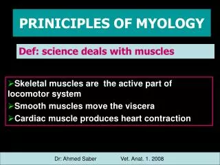
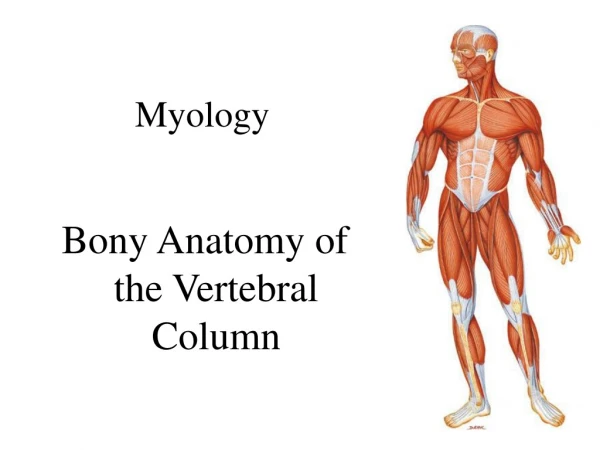
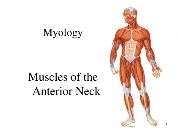
MYOLOGY STUDY OF MUSCLE IS CALLED “MYOLOGY”. MUSCLE : It is an ordinary arrangement of Connective tissue and contractile cells. FUNCTIONS : 1. Movement -> alternative contraction and relaxation

E N D
MYOLOGYSTUDY OF MUSCLE IS CALLED “MYOLOGY” MUSCLE : It is an ordinary arrangement of Connective tissue and contractile cells. FUNCTIONS : 1. Movement -> alternative contraction and relaxation 2. Protection of vessels and nerves 3. Pumping of blood. 5. Shapes to the organs & body 6. Transportation (food, blood, waste products)
MYOLOGY • TYPES OF MUSCLES : - Based on their shape and innervation muscles are divided in to 3 Types 1. SKELETAL MUSCLE (Striated Voluntary) 2. CARDIAC MUSCLE (Striated Involuntary) 3. SMOOTH MUSCLE (Plain Involuntary)
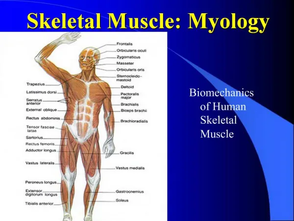
MYOLOGY 1. SKELETAL MUSCLE :- • Attached to bone and produce movement. • Also called striated voluntary. (Presence of striations and under will control) PARTS (MACROSTRUCTURE) :- • PROXIMAL ATTACHEMENT (ORIGIN OR HEAD) 2. BELLY (BODY) 3. DISTAL ATTACHEMENT (INSERTION OR TENDON)
MYOLOGY 1. PROXIMAL ATTACHEMENT :- Attachment that least moves 2. DISTAL ATTACHEMENT :- Attachment that moves most - Ends of muscles form in to cards of fiber tissue called “TENDONS” 3. Belly :- - Middle flabby part of muscle is called “BELLY” APONEUROSIS:- Thin & strong fibrous sheet of flat muscles RAPHAE:-Inter digitations of tendenious ends of muscle fibers
MYOLOGY • STRUCTURE OF SKELETAL MUSCLE (MICRO):- • PARTS :- 1. FASCICLE 2. MUSCLE CELL (Muscle fiber) 3. MYOFIBRIEL 4. MYOFILEMENTS
MYOLOGY 1.FASCICLE :- - Group of muscle cell bind together and form “Fascicle” 2. Muscle cell :- - Elongated cells 3. MYOFIBRIEL:- - Cylindrical, threadlike bundles of myofilaments 4. MYOFILAMENT:- - Contractile protein elements of muscle
MYOLOGY • MUSCLE CELL OR FIBER :- (INTRACELLULAR ORGANIZATION) 1. NUCLEUS : Cellular structure contains D.N.A. (genetic) material 2. SARCOLEMMA :Specific name of the plasma membrane. 3. SARCOPLASMIC RETICULUM (S.R.) : Name of Endoplasmic reticulum. It interconnects tubules surrounding each myofibril like a slaves of a swatter 4.TERMINAL CISTERNAE : Sac like regions of Sarcoplasmic Reticulm (S.R.). It carries Ca+ ions.
MYOLOGY 5.T – TUBULES: Invaginations of sarcolemma projecting deep in to muscle fibers called “T-tubules” opens out on to the surface of the Sarcolemma. 6. TRIAD :- -> Terminal cisternae It is a group of 3 units -- - -> T-tubule - -> Terminal cisternae 7. CYTOSOL : Intra cellular fluid. Organelles of cell are suspended init. 8. MITOCHONDRIA : A.T.P. Synthesizers, powerhouses of cell, suspended in cytosol.
MYOLOGY MYOFIBRIL • MYOFIBRIL : - Thread like Cylindrical bundles of contractile filaments • MYOFILAMENTS : - Contractile proteins of muscle cell. 2 TYPES : 1. THIN FILAMENT (Protein – ACTIN) 2. THICK FILAMENT (Protein – MYOSIN )
MYOLOGY COVERS OF MUSCLE TISSUE Muscle tissue is covered by 3 layers of connective tissue sheathes. They are, • Epimysium : Outermost sheath covers each muscle bulk. • Perimysium : Middle sheath that covers each fascicle. • Endomysium : Innermost sheath that covers each muscle fiber.
1. SKELETAL MUSCLE • HISTOLOGY : - Elongated cells - Visible striations - Multiple,peripheral nuclei. - Voluntary Eg: Biceps brachi
CARDIAC MUSCLE : - Branched cells - single central nuclei - visible strations - Involuntary PROPERTY –> SPONTANIOUS &RHYTHMICAL CONTRACTION Eg: Myocardium of heart.
SMOOTH MUSCLE • - Spindle shaped cells - Single central nucleus - No visible striations - Involuntary Eg: Stomach, Urinary bladder, Uterus.
MYOLOGY • TWO JOINT MUSCLE : Muscle which crosses two or more joints and performs two opposite actions at the two different joints are called “TWO JOINT MUSCLE” Eg.: Gastrocnemius contraction -> Flexion of knee & Ext. of foot (D.F.) APPLIED ANATOMY : Spasticity (Hypertonecity): Increased tone of the muscle Flacidity (Hypotonecity) : Decreased tone of the muscle Atrophy or wasting : Decreased size of muscle Hypertrophy : Increased size of muscle bulk Fatigue : Short lasting weakness of muscle - x -