Download
1 / 27
290 likes | 328 Views
Myology. Muscles of the Anterior Neck. Muscles of the Neck Overview. Muscle of neck are divided into two groups: Anterior Superficial (2) Hyoids Infrahyoids (4) Suprahyoids (4) Scalenes (3) Deep (4) Posterior Superficial (4) Deep (4)

E N D
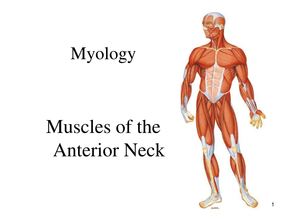
Myology Muscles of the Anterior Neck
Muscles of the Neck Overview • Muscle of neck are divided into two groups: • Anterior • Superficial (2) • Hyoids • Infrahyoids (4) • Suprahyoids (4) • Scalenes (3) • Deep (4) • Posterior • Superficial (4) • Deep (4) • Note: Some sources divide neck into anterior, posterior, & lateral.
Muscles of Neck Overview • Functionality • Since these muscles cross the joints of the cervical spine, they can move the neck at the cervical spinal joints • If a muscle also crosses the atlanto-occipital joint (C0/C1) then it can move the head upon the neck.
Muscles of Neck Overview • General Rules: • If a muscle crosses the neck posteriorly, it can extend the neck at the cervical spinal joints. • If a muscle crosses the neck anteriorly, it can flex the neck at the cervical spinal joints. • If a muscle crosses the neck laterally, it can laterally flex the neck at the cervical spinal joints. • If a muscle wraps around the neck, it can cause rotation of the neck at the cervical spinal joints.
Muscles of the Anterior Neck – Superficial (2) • Platysma : • By function it is primarily a muscle of facial expression i.e. innervated by CN VII. • Platysma of one side blends with contralateral side and other facial muscles in lower face. • Considered to by remnant of a broader muscle called panniculus carnosus found in four-legged animals. Enables horses to shake off flies and cats to raise hair on its back. • When contracted it is reminiscent of “Creature from the Black Lagoon” creature. • Sternocleidomastoid (SCM): • Since it attaches to sternum, SCM is considered an accessory muscle of respiration.
O: Subcutaneous Fascia of Superior Chest I: Mandible and subcutaneous fascia of lower face A: Draws up the skin of superior chest and neck, creating ridges in neck skin. Assists in drawing the lip laterally and depresses the mandible N: CN VII (Facial nerve) Platysma Palpation: Page 138
Sternocleidomastoid (SCM) O: Sternal Head: manubrium Clavicular Head: Medial clavicle I: Mastoid process Actions: Bilateral contraction: flexion of the neck. Unilateral contraction results in Lateral flexion of neck/head and Contralateral rotation of neck/head N: Spinal accessory nerve (CN XI) Palpation: Page 141
Muscles of the Anterior Neck – Infrahyoids (4) • All 4 infrahyoid muscles are located below the hyoid bone i.e. the pull hyoid bone inferiorly when contracted. • All hyoid muscle are important in moving and/or fixating the hyoid bone. These functions are necessary for chewing, swallowing, & speech. • Sternohyoid: • “Sterno” refers to sternum • “hyoid” refers to hyoid bone • Sternothyroid: • “thyroid” refers to thyroid cartilage • Thyrohyoid • Omohyoid: • “Omo” refers to the shoulder
Sternohyoid O: Posterior aspect of the manubrium and medial clavicle I: Inferior Hyoid A: Depression of hyoid N: Ansa cervicalis of the cervical plexus Palpation: page 147
Sternothyroid O: Posterior Sternum and 1st costal cartilage I: Thyroid Cartilage A: Depression of thyroid cartilage N: Ansa cervicalis of the cervical plexus Palpation: page 150
Thyrohyoid O: Thyroid Cartilage I: Hyoid (inferior aspect) A: Depression of hyoid and Elevation of thyroid cartilage N: CN XII (Hypoglossal nerve) Palpation: page 152
Omohyoid O: Inferior Belly: Superior angle of the scapula Superior Belly: Clavicle via the central bound to the clavicle I: Inferior belly: Clavicle (via the central bound to the clavicle) Superior belly: hyoid A: Depression of hyoid N: Ansa cervicalis of the cervical plexus Palpation: page 155
Muscles of the Anterior Neck – Suprahyoids (4) • Digastric: • “Di” means two; “gastric” means belly • External carotid lies inferior and deep to anterior belly • Stylohyoid: • External carotid lies inferior and deep to stylohyoid • Mylohyoid: • “mill” refers to molar teeth • Geniohyoid: • “genio” refers to chin
Digastric O: Posterior belly: mastoid notch of temporal bone Anterior belly: Inner surface of the mandible I: Hyoid (via the central tendon) A: Elevation of hyoid, depression of the mandible, and retraction of the mandible. N: anterior belly: CN V (Trigeminal nerve) posterior belly CN VII (Facial nerve) Palpation: page 158
Stylohyoid O: Styloid process of temporal bone I: Hyoid Actions: Elevation of hyoid N: CN VII (Facial nerve) Palpation: page 161
Mylohyoid O: Entire inner surface of mandible (this muscle forms the muscular floor of the mouth) I: Hyoid A: Elevation of hyoid and depresses the mandible N: CN V (Trigeminal nerve) Palpation: page 164
Geniohyoid O: Inner surface of mandible, deep to the mylohyoid I: Hyoid A: Elevation of hyoid N: CN XII (Hypoglossal nerve) Palpation: page 167
Muscles of the Anterior Neck – Scalenes (3) • As a group, they attach superiorly from cervical TP's to inferiorly on the 1st and 2nd ribs • As a group, scalenes flex and laterally flex the neck • By reverse muscles action, the scalenes can elevated the 1st & 2nd rib i.e. they are also considered accessory muscle of respiration.
Anterior Scalene O: Anterior tubercles of the TP’s of C3 – C6 I: 1st Rib A: Bilateral contraction: flexion of the neck. Unilateral contraction causes lateral flexion and contralateral rotation of the neck. Reversed muscle action causes Elevation of 1st rib N: Ventral rami of the cervical spinal nerves Palpation: page 173
Middle Scalene O: Posterior tubercles of the TP’s of C2 to C7 I: 1st Rib A: Bilateral contraction: flexion of the neck. Unilateral contraction causes lateral flexion of the neck. Reversed muscle action causes Elevation of 1st rib N: Ventral rami of the cervical spinal nerves Palpation: page 176
Posterior Scalene O: Posterior tubercles of the TP’s of cervical spine I: 2nd Rib A: Unilateral contraction causes lateral flexion of the neck. Reversed muscle action causes Elevation of 2nd rib N: Ventral rami of the cervical spinal nerves Palpation: page 179
The Scalene Group • Scalenes, as well as SCM, are often injured during MVA called whiplash. • Also known as cervical acceleration deceleration (CAD) injury
Muscles of the Anterior Neck – Deep Prevertebral Group (4) • Called prevertebral muscles since they lie directly on the cervical spine vertebral bodies • Important at fixating (stabilizing) and neck/head while talking, swallowing, coughing, & sneezing • May also be injured during CAD • Longus Colli: • Has 3 parts: superior oblique, inferior oblique, & vertical • Considered to be a strong neck flexor • Longus Capitis • Rectus Capitis Anterior • Rectus Capitis Lateralis
Longus Colli O: Bodies of the C3-T3 vertebrae I: TP’s and Bodies of the C1-C6 vertebrae A: Bilateral contraction causes weak flexion of neck. Unilateral contraction causes lateral flexion and contralateral rotation of the neck. N: Ventral rami of the cervical spinal nerves For the purpose of HS 113, this muscle is not palpable
Longus Capitis O: TP’s of C3 – C5 I: Basilar portion of the occiput A: Bilateral contraction causes Flexion of head/neck. Unilateral contraction causes Lateral flexion of head/neck N: Ventral rami of the cervical spinal nerves For the purpose of HS 113, this muscle is not palpable
Rectus Capitis Anterior O: TP of the Atlas (C1) I: Inferior surface of the basilar portion of the occiput A: Flexion of head N: Ventral rami of the cervical spinal nerves For the purpose of HS 113, this muscle is not palpable
Rectus Capitis Lateralis O: TP of the Atlas (C1) I: Inferior surface of the Occiput A: Lateral flexion of head N: Ventral rami of the cervical spinal nerves For the purpose of HS 113, this muscle is not palpable