Download
1 / 11
110 likes | 117 Views
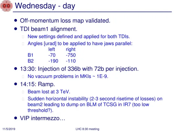
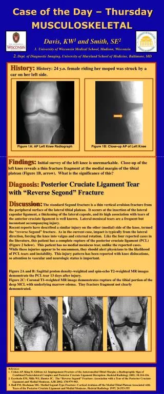
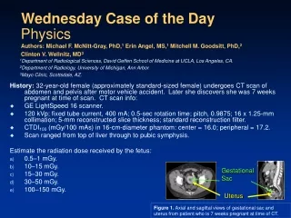
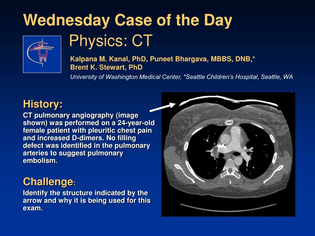
History: CT pulmonary angiography (image shown) was performed on a 24-year-old female patient with pleuritic chest pain and increased D-dimers. No filling defect was identified in the pulmonary arteries to suggest pulmonary embolism. Challenge :

E N D
History: CT pulmonary angiography (image shown) was performed on a 24-year-old female patient with pleuritic chest pain and increased D-dimers. No filling defect was identified in the pulmonary arteries to suggest pulmonary embolism. Challenge: Identify the structure indicated by the arrow and why it is being used for this exam. Wednesday Case of the Day Physics: CT Kalpana M. Kanal, PhD, Puneet Bhargava, MBBS, DNB,* Brent K. Stewart, PhD University of Washington Medical Center, *Seattle Children’s Hospital, Seattle, WA
Answer : The structure identified by the arrow is an image of a bismuth shield used during the clinical exam. Its purpose is to reduce the dose to the breast of the 24-year-old female patient. Wednesday Case of the Day Physics: CT Kalpana M. Kanal, PhD, Puneet Bhargava, MBBS, DNB,*Brent K. Stewart, PhD University of Washington Medical Center, *Seattle Children’s Hospital, Seattle, WA
Discussion What is the breast dose from CT? The breast is a radiosensitive organ. The dose to the breast from a CT PE examination is estimated to be 20-60 mGy, the dose for a CT coronary angiography examination is estimated to be 50-80 mGy, and the dose to the inferior part of the breast for an abdominal CT examination is estimated to be 10-20 mGy (Mettler et al). In comparison, a two-view mammogram imparts an average dose of 2 mGy to the breast (Bushberg et al). Wednesday Case of the Day Physics: CT Kalpana M. Kanal, PhD, Puneet Bhargava, MBBS, DNB,* Brent K. Stewart, PhD University of Washington Medical Center, *Seattle Children’s Hospital, Seattle, WA
Discussion What is the breast dose from CT? Figure shows the radiation dose to the breast for a PE protocol using a multidetector CT scanner. Dose to breasts ranged from 35-42 mGy. (Reprinted, with permission, from Hurwitz et al.) Wednesday Case of the Day Physics: CT Kalpana M. Kanal, PhD, Puneet Bhargava, MBBS, DNB,*Brent K. Stewart, PhD University of Washington Medical Center, *Seattle Children’s Hospital, Seattle, WA
Discussion What is the breast cancer risk from CT? The graph shows the % lifetime attributable risk of breast cancer incidence from a single standard CT coronary angiography exam (would be similar for CT PE exam). The risk is higher for younger women and decreases with age. Lifetime Attributable Risk of Cancer Incidence % Wednesday Case of the Day Physics: CT Kalpana M. Kanal, PhD, Puneet Bhargava, MBBS, DNB,*Brent K. Stewart, PhD University of Washington Medical Center, *Seattle Children’s Hospital, Seattle, WA Einstein et al, 2007.
Discussion What is the breast cancer risk from CT? The table shows that the lifetime attributable risk (per 100,000 exposed people) of breast cancer for a 25-year-old who underwent a PE exam is 133, compared to 20 for a 55-year-old. (Reprinted, with permission, from Hurwitz et al.) Wednesday Case of the Day Physics: CT Kalpana M. Kanal, PhD, Puneet Bhargava, MBBS, DNB,*Brent K. Stewart, PhD University of Washington Medical Center, *Seattle Children’s Hospital, Seattle, WA
Discussion What can we do to reduce dose to the female breast? Consider and, if possible, use alternative imaging techniques such as US and MRI to avoid radiation exposure to the breast altogether. Limit the field of view, if possible, to minimize the amount of area irradiated. For example, there is no need to include most of the lower chest in an abdominal CT study being performed for evaluation of right-lower-quadrant abdominal pain. Alter scan parameters (eg, low-dose technique using low kVp or mAs for follow-up scans of pulmonary nodules). Wednesday Case of the Day Physics: CT Kalpana M. Kanal, PhD, Puneet Bhargava, MBBS, DNB,*Brent K. Stewart, PhD University of Washington Medical Center, *Seattle Children’s Hospital, Seattle, WA
Discussion What can we do to reduce dose to the female breast? Avoid multiphase acquisition when not necessary (eg, for most studies, noncontrast CT images are not necessary if postcontrast CT is being performed). Follow recommended follow-up guidelines. For example, follow Fleischner Society* guidelines for small pulmonary nodules and to decrease cumulative radiation exposure by delaying follow-up if the patient is low risk. Use bismuth shields to protect the breasts of young female patients. *MacMahon H, Austin JHM, Gamsu G, et al. Guidelines for management of small pulmonary nodules detected on CT scans: a statement from the Fleischner Society. Radiology 2005;237:395-400. Wednesday Case of the Day Physics: CT Kalpana M. Kanal, PhD, Puneet Bhargava, MBBS, DNB,*Brent K. Stewart, PhD University of Washington Medical Center, *Seattle Children’s Hospital, Seattle, WA
Discussion What can we do to reduce dose to the female breast? Bismuth shielding (arrow) is effective in reducing dose to the breast. At our institution, we have seen a decrease of 37% in the breast dose when using bismuth shields, without significant degradation in image quality. Wednesday Case of the Day Physics: CT Kalpana M. Kanal, PhD, Puneet Bhargava, MBBS, DNB,*Brent K. Stewart, PhD University of Washington Medical Center, *Seattle Children’s Hospital, Seattle, WA
Discussion What can we do to reduce dose to the female breast? Fricke et al showed a 29% reduction in breast dose by using bismuth shields on pediatric patients, without any significant change in image quality. Hohl et al showed a 32% breast dose reduction using bismuth shields, without deterioration in image quality. Wednesday Case of the Day Physics: CT Kalpana M. Kanal, PhD, Puneet Bhargava, MBBS, DNB,*Brent K. Stewart, PhD University of Washington Medical Center, *Seattle Children’s Hospital, Seattle, WA
References/Bibliography Einstein et al. JAMA, July 18, 2007 – Vol. 298, No. 3, page 317. Hurwitz et al. Radiology, December 2007 – Vol. 245, No. 3, page 742. Mettler et al. Radiology, July 2008 – Vol. 248, No. 1, page 254. Fricke et al. AJR, February 2003 – Vol. 180, page 407. Bushberg et al. The Essential Physics of Medical Imaging, 2nd edition, 2002. Hohl et al. Acta Radiologica, March 2006 – Vol. 27, No. 6, page 562. http://radiology.rsnajnls.org/cgi/content/full/237/2/395 Wednesday Case of the Day Physics: CT Kalpana M. Kanal, PhD, Puneet Bhargava, MBBS, DNB,*Brent K. Stewart, PhD University of Washington Medical Center, *Seattle Children’s Hospital, Seattle, WA