Download
1 / 7
70 likes | 77 Views
This case study examines the impact of a highly attenuating object on a pediatric CT scan. It discusses the effects on patient dose, image noise, and beam hardening artifact.

E N D
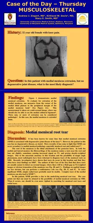
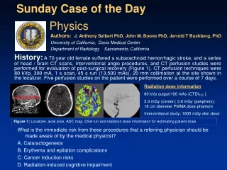
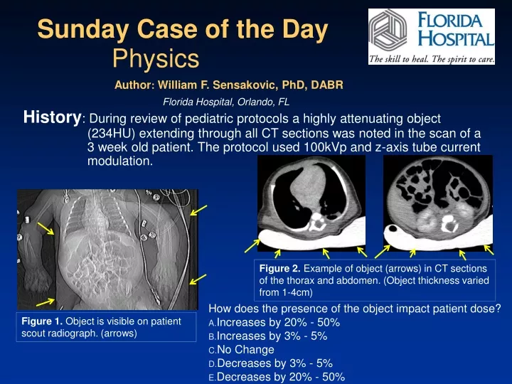
History: During review of pediatric protocols a highly attenuating object (234HU) extending through all CT sections was noted in the scan of a 3 week old patient. The protocol used 100kVp and z-axis tube current modulation. Sunday Case of the Day Physics • How does the presence of the object impact patient dose? • Increases by 20% - 50% • Increases by 3% - 5% • No Change • Decreases by 3% - 5% • Decreases by 20% - 50% Author: William F. Sensakovic, PhD, DABR Florida Hospital, Orlando, FL Figure 2. Example of object (arrows) in CT sections of the thorax and abdomen. (Object thickness varied from 1-4cm) Figure 1. Object is visible on patient scout radiograph. (arrows)
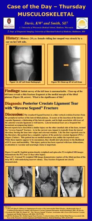
A solid water phantom was scanned with and without the warming mattress in the CT scanners of three different manufacturers. (Figs. 3&4) Tube current selection, tube current modulation, and tube voltage selection were also varied when available for each CT scanner. Findings:Discussions with CT technologists and pediatric nursing revealed that the object was a warming mattress used to prevent hypothermia in neonates Figure 3: Phantom with warming mattress Figure 4: Phantom without warming mattress
Noise (standard deviation) was increased in regions of interest placed at the posterior level of the warming mattress. This occurred even when tube current modulation and tube voltage selection were implemented. (Figs. 5&6) Mean HU was decreased in regions of interest placed at the anteroposterior level of the warming mattress. This occurred even when tube current modulation and tube voltage selection were implemented. (Figs. 5&6) Findings: (continued) Figure 5: No warmer, 100kVp, Automated tube current selection implemented Mean: 8.0HU Standard deviation: 13.3HU Figure 6: Same parameters with warmer causing increased noise and beam hardening Mean: 0.2HU Standard deviation: 29.3HU
At diagnostic x-ray energies, the radiation is roughly halved every 3.5cm of soft tissue the beam travels through. The more attenuation the CT scanner expects in the path of the radiation beam, the higher the mAs and/or kVp are set. The pediatric warming mattress is several centimeters thick and has an attenuation coefficient between water and bone (as is apparent from its HU value). The CT scanners tested increased the tube current 20% - 50% to compensate for the increased attenuation of the warming mattress. Despite the increased mAs generated by the machine when the pediatric warming mattress is used, increased image noise and decreased mean HU (beam hardening artifact ) are still observed at the posterior level of the warming mattress. (Fig. 6) Discussion: Automated techniques to select and modulate mAs and automated techniques to select kVp are generally implemented to reduce patient dose. The CT scanner determines the mAs/kVp to apply by analyzing the pre-scan planning radiograph (Fig. 7) and/or the radiation detected from previous sections in the current CT scan. Figure 7: Pre-scan planning radiographs without (top) and with (bottom) pediatric warming mattress. Increased attenuation from the warming mattress causes the CT scanner to increase patient dose.
Discussion: Although it is theoretically possible that the presence of the warming mattress could cause the machine to select a higher kVp when automated tube voltage selection is implemented, this was not observed in phantom experiments. Some scans using angular tube current modulation demonstrated relatively small increases in patient dose, noise, and beam hardening artifact compared to scans using tube current selection alone. This was vendor specific and differences were likely due to different algorithms used to control modulation. Figure 8: Phantom images with pediatric warming mattress in place. Left: Image with angular dose modulation implemented. Right: Image with automatic tube current selection, but not angular modulation
References/Bibliography: Mannudeep KK, et al. Techniques and Applications of Automatic Tube Current Modulation for CT. Radiology 233(3): 649, 2004. Raman SP, et al. CT Dose Reduction Applications: Available Tools on the Latest Generation of CT Scanners.J Am Coll Radiol 10: 37, 2013. Siegel MJ, et al. Automated low-kilovoltage selection in pediatric computed tomography angiography: phantom study evaluating effects on radiation dose and image quality. Invest Radiol 48(8): 584, 2013.