Download
1 / 71
710 likes | 1.32k Views
Class 6 (Initial Assessment with Circulatory System, Bleeding & Shock) Ch8 (Partial) , Ch4 (Partial) , Ch22 & Ch23. Patient Assessment (Review). Scene size-up Initial assessment Focused history and physical exam Vital signs History Detailed physical exam Ongoing assessment.

E N D
Class 6 (Initial Assessment with Circulatory System, Bleeding & Shock)Ch8 (Partial), Ch4 (Partial), Ch22 & Ch23
Patient Assessment (Review) • Scene size-up • Initial assessment • Focused history and physical exam • Vital signs • History • Detailed physical exam • Ongoing assessment
Initial Assessment • Develop a general impression. • Assess mental status. • Assess airway. • Assess the adequacy of breathing. • Assess circulation. • Identify patient priority.
Initial Assessment: A-B-C Triad C • Find & treat immediate threat to life • Lack of an airway • Ineffective breathing • Circulatory collapse • Bleeding • Shock • A B C all of equal importance • Starting with bleeding control (C) appropriate in an awake patient A B
Anatomy of theCardiovascular System • The cardiovascular system is responsible for supplying and maintaining adequate blood supply flow. • Consists of three parts: • Heart (pump) • Blood vessels (container) • Blood and body fluids (fluids)
Blood Vessels • Arteries • Arterioles • Capillaries • Venules • Veins
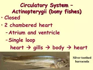
Aorta Pulmonary Carotid Femoral Brachial Radial Superior vena cava Inferior vena cava Pulmonary Major Arteries and Veins
Capillary Sphincters • Regulate the blood flow through the capillary beds. • Sphincters are under the control of the automatic nervous system. • Regulation of blood flow is determined by cellular need.
Blood • Contains: • Red blood cells • White blood cells • Platelets • Plasma
Physiology of theCirculatory System (1 of 2) • Pulse • The wave of blood through the arteries formed when the left ventricle contracts • Can be felt where an artery passes near the skin surface and over a bone
Physiology of theCirculatory System (2 of 2) • Blood pressure • Amount of force exerted against walls of arteries • Systole: Left ventricle contracts • Diastole: Left ventricle relaxes • Perfusion • Circulation of blood within an organ or tissue • If inadequate, the patient goes into shock.
Perfusion (1 of 2) • Circulation within tissues in adequate amounts to meet the cells’ needs for oxygen, nutrients, and waste removal • Some tissues and organs need a constant supply of blood while others can survive on very little when at rest.
Perfusion (2 of 2) • The heart demands a constant supply of blood. • The brain and spinal cord can survive for 4 to 6 minutes without perfusion. • The kidneys may survive 45 minutes. • The skeletal muscles may last 2 hours.
Perfusion Triangle Blood Vessels (Container Function) Heart (Pump Function) Blood (Content Function)
C for Circulation, Bleeding & Shock
Assessing the Pulse • Presence • Rate • Rhythm • Strength
The Significance of Bleeding • Hemorrhage = bleeding • The body will not tolerate an acute blood loss of greater than 20% of the blood volume. • In the typical adult, 20% is 1 liter or 2 pints. • Blood loss of 1 L can be dangerous in adults; in children, loss of 100-200 mL. is serious • A 1-year-old infant typically has 800 mL. A loss of 200 mL is significant.
Conditions With Possible Serious Bleeding • Significant mechanism of injury • Poor general appearance of patient • Assessment reveals signs of shock • Significant amount of blood loss noted • Blood loss is rapid. • You cannot control external bleeding.
Assessing and Controlling External Bleeding • Assess after clearing the airway and stabilizing breathing. • Look for blood flow or blood on floor/clothes. • Controlling bleeding • Direct pressure • Elevation • Pressure points
Characteristics of Bleeding • Arterial • Blood is bright red and spurts. • Venous • Blood is dark red and does not spurt. • Capillary • Blood oozes out and is controlled easily.
Blood Clotting • Bleeding normally stops within 10 minutes. • Some medications interfere with clotting. • Some injuries will be unable to clot. • Patients with hemophilia lack clotting factors.
Circulation • Quickly assess pulse rate and quality. • Determine skin condition, color, and temperature. • Control significant bleeding. • Treat for shock.
Interventions • With significant bleeding, provide high-flow oxygen. • Control bleeding. • Using multiple methods to control bleeding usually works best. • Treat aggressively for shock. • Provide rapid transport.
Controlling External Bleeding • Follow BSI precautions. • Ensure patient has an open airway and adequate breathing. • Provide oxygen if necessary. • There are several methods to control bleeding. • Direct pressure & elevation • Pressure points • Splints • Tourniquets
Direct Pressure and Elevation • Direct pressure is the most common and effective way to control bleeding. • Apply pressure with gloved finger or hand. • Elevating a bleeding extremity often stops venous bleeding. • Use both direct pressure and elevation whenever possible. • Apply a pressure dressing.
Pressure Points • If bleeding continues, apply pressure on pressure point. • Pressure points are located where a blood vessel lies near a bone. • Be familiar with the location of pressure points.
Splints • Splints can help control bleeding associated with a fracture. • Air splints can be used to control bleeding of soft-tissue injuries.
Applying a Tourniquet • Fold a triangular bandage into 4˝ cravat. • Wrap the bandage. • Use a stick as a handle to twist and secure the stick. • Write “TK” and time. Place on patient.
Tourniquet Precautions • Place as close to injury as possible, but not over joint. • Never use narrow material. • Use wide padding under the tourniquet. • Never cover a tourniquet with a bandage. • Do not loosen the tourniquet once applied.
Bleeding from the Nose,Ears, and Mouth • Causes: • Skull fractures • Facial injuries • Sinusitis • High blood pressure • Coagulation disorders • Digital trauma
Controlling a Nosebleed • Follow BSI precautions. • Help the patient sit and lean forward. • Apply direct pressure by pinching the patient’s nostrils. • Or place a piece of gauze bandage under the patient’s upper lip and gum. • Apply ice over the nose. • Provide transport.
Bleeding from Skull Fractures • Do not attempt to stop the blood flow. • Loosely cover bleeding site with sterile gauze. • If cerebrospinal fluid is present, a target (or halo) sign will be apparent.
Internal Bleeding • Internal bleeding may not be readily apparent. • Assess patient’s: • Mechanism of injury • Nature of illness
Signs and Symptomsof Internal Bleeding (1 of 2) • Ecchymosis: Bruising • Hematoma: Bleeding beneath the skin • Hematemesis: Blood in vomit • Melena: Black, tarry stool
Signs and Symptomsof Internal Bleeding (2 of 2) • Hemoptysis: Coughing up blood • Pain, tenderness, bruising, guarding, or swelling • Broken ribs, bruises over the lower chest, or rigid, distended abdomen • Referred pain
Shock Shock = Inadequate Tissue Perfusion • State of collapse and failure of the cardiovascular system • Leads to inadequate circulation • Without adequate blood flow, cells cannot get rid of metabolic wastes • The result of hypoperfusion to cells that causes the organ, then organ systems, to fail
Perfusion Triangle Blood Vessels (Container Function) If all the vessels dilate at once, the normal amount of blood volume is not enough to fill the system and provide adequate perfusion to the body. Heart (Pump Function) Damage to the heart by disease or injury. It cannot move blood adequately to support perfusion. Blood (Content Function) If blood or plasma is lost, the volume in the container is not enough to support the perfusion needs of the body.
Change in mental status Tachycardia Weakness Thirst Nausea or vomiting Cold, moist skin Shallow, rapid breathing Dull eyes Dilated pupils Weak, rapid pulse Decreased blood pressure Altered level of consciousness Signs of Hypoperfusion
Progression of Shock • Compensated shock • When the body compensates for blood loss • Decompensated shock • The late stage of shock when blood pressure is falling • Irreversible shock • The terminal stage
Agitation Anxiety Restlessness Feeling of impending doom Altered mental status Weak pulse Clammy skin Pallor Shallow, rapid breathing Shortness of breath Nausea or vomiting Delayed capillary refill Marked thirst Compensated Shock
Decompensated Shock • Falling blood pressure (<90 mm Hg in an adult) • Labored, irregular breathing • Ashen, mottled, cyanotic skin • Thready or absent pulse • Dull eyes, dilated pupils • Poor urinary output
Irreversible Shock • This is the terminal stage of shock. • A transfusion of any type will not be enough to save a patient’s life.
When to Expect Shock • Multiple severe fractures • Abdominal or chest injuries • Spinal injuries • Severe infection • Major heart attack • Anaphylaxis
Cardiovascular Causes of Shock (1 of 4) • Pump failure (cardiogenic shock) • Inadequate function of the heart or pump failure • Causes a backup of blood into the lungs • Results in pulmonary edema • Pulmonary edema leads to impaired ventilation
Cardiovascular Causes of Shock (2 of 4) • Poor vessel function (neurogenic shock) • Damage to the cervical spine may affect control of the size and muscular tone of blood vessels. • The vascular system increases. • Blood in the body cannot fill the enlarged system. • Neurogenic shock occurs.