Download
1 / 41
410 likes | 611 Views
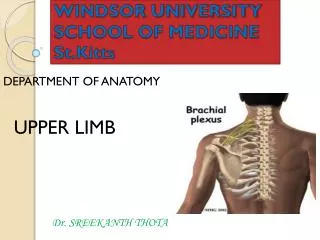
WINDSOR UNIVERSITY SCHOOL OF MEDICINE St.Kitts. Dr. SREEKANTH THOTA. DEPARTMENT OF ANATOMY. UPPER LIMB. FOREARM . FOREARM. The forearm is the part of the upper limb that extends between the elbow joint and the wrist joint.

E N D
WINDSOR UNIVERSITYSCHOOL OF MEDICINESt.Kitts Dr. SREEKANTH THOTA DEPARTMENT OF ANATOMY UPPER LIMB FOREARM
FOREARM • The forearm is the part of the upper limb that extends between the elbow joint and the wrist joint. • As in the arm, the forearm is divided into anterior and posterior compartments
Compartments of forearm • Muscles in the anterior compartment of the forearm flex the wrist and digits and pronate the hand. • Muscles in the posterior compartment extend the wrist and digits and supinate the hand.
ANTERIOR COMPARTMENT OF THE FOREARM • Muscles in the anterior (flexor) compartment of the forearm occur in three layers: • 1. Superficial • 2. Intermediate • 3.Deep • All muscles in the anterior compartment of the forearm are innervated by the median nerve, except for the flexor carpi ulnaris muscle and the medial half of the flexor digitorum profundus muscle, which are innervated by the ulnar nerve.
Superficial layer • Four muscles in the superficial layer- • 1.flexor carpi ulnaris • 2.palmaris longus • 3.flexor carpi radialis • 4. pronator teres- • All four muscles have a common origin from the medial epicondyle of the humerus, and, except for pronator teres, extend distally from the forearm into the hand
Intermediate layer • Flexor digitorum superficialis
Deep layer • 1.flexor digitorum profundus • 2.flexor pollicis longus • 3.pronator quadratus
Nerves • Nerves in the anterior compartment of the forearm are the median and ulnar nerves, and the superficial branch of the radial nerve.
Median nerve • The median nerve innervates the muscles in the anterior compartment of the forearm except for the flexor carpi ulnaris and the medial part of the flexor digitorum profundus (ring and little fingers). • It leaves the forearm and enters the palm of the hand by passing through the carpal tunnel deep to the flexor retinaculum.
Branches of median nerve • 1. Anterior interosseous nerve: innervates the muscles in the deep layer • 2. A small palmar branchoriginates from the median nerve in the distal forearm immediately proximal to the flexor retinaculum. • Innervates the skin over the base and central palm. • This palmar branch is spared in carpal tunnel syndrome because it passes into the hand superficial to the flexor retinaculum of the wrist.
Ulnar nerve • In the forearm, the ulnar nerve innervates only the flexor carpi ulnaris muscle and the medial part (ring and little fingers) of the flexor digitorum profundus muscle. • Branches in forearm: • 1.Palmar branchoriginates in the middle of the forearm and passes into the hand to supply skin on the medial side of the palm
POSTERIOR COMPARTMENT OF THE FOREARM • Muscles in the posterior compartment of the forearm occur in two layers: • 1.Superficial • 2.Deep layer • The muscles are associated with: • 1.Movement of the wrist joint • 2.Extension of the fingers and thumb • 3.Supination. • All muscles in the posterior compartment of the forearm are innervated by the radial nerve.
Superficial layer • Seven muscles • 1.Brachioradialis • 2.Extensor carpi radialis longus • 3.Extensor carpi radialis brevis • 4.Extensor digitorum • 5.Extensor digiti minimi • 6.Extensor carpi ulnaris • 7.Anconeus
Brachioradialis • Action • Flexes forearm
Extensor carpiradialislongus and brevis • Action • Extends and abducts hand at wrist joint
Extensor digitorum • Action • Extends the medial four digits at metacarpo –phalangeal, IP joint and extends hand at wrist
Extensor digitiminimi • Action • Extends the 5th digit at the metacarpal-phalangeal and interphalangeal joints
Extensor carpiulnaris • Action • Extends and adducts hand at the wrist
Anconeus • Action • accessory extensor of the elbow joint
Deep layer • Five muscles • 1-Supinator • 2-Abductor pollicis longus • 3-Extensor pollicis brevis • 4-Extensor pollicis longus • 5-Extensor indicis
Supinator • Action • Supinates forearm
Abductor pollicislongus • Action • Abducts thumb
Extensor pollicislongus and brevis • Action • 1. Extensor Pollicis Longus • Extends distal phalanx of thumb • 2. Extensor Pollicis Brevis • Extends proximal phalanx of thumb
Extensor indicis • Action • Extends index finger
ANATOMICAL SNUFFBOX • Boundries • Medially • -Extensor pollicislongus tendon • Laterally • Abductor pollicislongus tendon • Extensor pollicisbrevis tendon • Clinical importance • 1- Scaphoid bone • 2- Radial pulsation
Radial nerve • The nerve of the posterior compartment of the forearm is the radial nerve . • In the lateral wall of the cubital fossa, and before dividing into superficialand deep branches, the radial nerve innervates the brachioradialis and extensor carpi radialis longus muscles. • Posterior interosseous nerve supplies the remaining muscles in the posterior compartment.
Superficial branch of Radial nerve The superficial branchof the radial nerve is sensory.
Radial Nerve Injury in the Forearm • Injury to the deep branch of the radial nerve may occur when wounds of the forearm are deep (penetrating). Severance of the deep branch of the radial nerve results in an inability to extend the thumb and the metacarpophalangeal (MP) joints of the other digits.
Testing the radial nerve • Integrity of the deep radial nerve may be tested by asking the person to extend the MP joints while the examiner provides resistance