Download
1 / 108
1.22k likes | 1.61k Views
Acute stroke imaging and endovascular therapy. mCgILL Neurology Academic half-day Wednesday, may 18 th 2011 Alexandre Poppe MD CM, FRCPC Hopital Notre-Dame, CHUM. Outline. Introduction CT or MRI? Parenchyma Vessels Perfusion Some cases

E N D
Acute stroke imagingand endovascular therapy mCgILL Neurology Academic half-day Wednesday, may 18th 2011 AlexandrePoppe MD CM, FRCPC Hopital Notre-Dame, CHUM
Outline Introduction CT or MRI? Parenchyma Vessels Perfusion Some cases Endovascular treatment of acute ischaemic stroke
Acute stroke imaging: the goals • Effectiveness of AIS therapy (e.g. thrombolysis) is time-dependent and requires rapid, accurate diagnosis • Imaging is essential to make a CORRECT diagnosis • R/O ICH • R/O stroke mimics (tumours, SDH etc.) • Clinical features do not reliably differentiate AIS from ICH
ICH or AIS? 55 y.o. male with acute left hemiparesis, N/V and headache Courtesy K. Butcher
CT or MRI? bmj.com aspectsinstroke.com
The ideal brain imaging technique Widely available Inexpensive Not harmful Fast Easy access to patient Differentiate AIS from ICH and mimics Provide good anatomical resolution Identify irreparably damaged tissue from salvageable tissue Adapted from Stroke: practical management. 3rd ed. 2007
Computed tomography (CT) • Clinical use for almost 40 years • Axial images by spiral acquisition using x-rays • 0.5 – 1.0 cm (anterior-middle fossa) • 0.25-0.5 cm (posterior fossa) • Image acquisition in about 10 seconds • CT angiography (CTA) requires iodinated contrast • Non-contrast CT (NCCT): radiation equivalent of about 150 chest x-rays
Magnetic resonance imaging (MRI) • In clinical use since the early 1980’s • Limited use in acute stroke until the last 10 years • No radiation • Routine MRI stroke protocol should include: • DWI: cytotoxicedema • FLAIR and T2: brain pathology (vasogenic, cytotoxicedema) • GRE: hemoglobin breakdown products (acute and remote bleeds) • T1: brain anatomy
Multimodal CT Advantages Disadvantages Widely-available Rapidly accessible Less expensive Short scanning times Few contraindications Excellent for exclusion of ICH CTA and CTP possible with iodinated contrast Lower sensitivity for acute ischemia (esp. small volume infarcts) Radiation exposure Contrast allergy and nephropathy Limited anatomical coverage for CTP
Multimodal MRI Advantages Disadvantages Excellent sensitivity for acute ischemia Reliable exclusion of ICH and stroke mimics Vessel and perfusion imaging possible with gadolinium Less available Longer scanning times More expensive Physically difficult for acutely-ill patients (1/3 require intervention during scan) 15-20% of acute stroke patients unable to undergo MRI
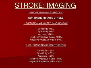
CT vs MRI: acute infarct detection • MRI (DWI) superior to NCCT for detection of AIS <12hrs, n=221 (OR 25 95%CI 8-79)1 • CT sens 16%, spec 97% • MRI sens 78%, spec 96% • Similar results for subgroup <3 hrs (n=90) • Interrater reliability better for MRI (kappa 0.84) vs CT (kappa 0.51)2 • False-negative DWI in • Posterior circulation AIS • Clinically mild stroke (NIHSS <4) • With GRE, MRI = CT for ICH detection Chalela et al. Lancet 2007 Fiebach et al. Stroke 2002
CT vs MRI: acute infarct detection • MRI useful for • Small subcortical infarcts • Brainstem infarcts • Small cortical infarcts (isolated or embolic shower) • DWI can differentiate acute from chronic lesions
Isolated cortical infarcts “right cortical hand”
However... CT remains the modality of choice at most Canadian stroke centres
The ischemic penumbra1 www.radiologyassistant.nl 1.Astrup Stroke 1981 • Acute arterial occlusion reduced CBF • Infarct core: • CBF too low to sustain cellular membrane integrity (ion pump failure) • <10ml/100g/min • Tissue death within minutes • Ischemic penumbra: • CBF too low to maintain electrical activity, but enough to maintain membrane integrity (10-20ml/100g/min) • Potentially salvageable tissue Courtesy K. Butcher Kidwell C
The ischemic penumbra • Penumbral tissue: • hypoperfused, hypoxic but structurally intact • At risk for infarction if perfusion not restored (time-dependent) • Current acute stroke therapies aim to prevent conversion of penumbral tissue into infarcted tissue • Restoring perfusion (CBF) by recanalizing AOL • “Buying time” to recanalize AOL by augmenting collateral circulation • Limiting recruitment of penumbra into core using “neuroprotection”
Le saint graal... Can imaging help us select those patients who are the best candidates for reperfusion therapy? And conversely exclude those with nothing to gain/at high risk for hemorrhage? Help guide which therapy to use? Help with prognostication?
What should we image? • Parenchyma • NCCT • MRI (DWI, FLAIR, T2) • Vessels • CTA • MRA • TCD • Perfusion (?penumbra) • CTP • MRI-PWI
Parenchymal imaging: CT • Identifies areas of recent infarction as • Hypoattenuation (reflects increased tissue water) • Loss of grey-white matter differentiation • Sulcal effacement/local swelling or mass effect Subacute infarct (>24 hours) Courtesy K. Butcher
Parenchymal imaging: CT Early ischemic changes (EICs) • Insular ribbon • ICA terminus occlusion, proximal and distal M1 occlusion • Lentiform nucleus • ICA terminus occlusion, proximal M1 occlusion • Corical ribbon • Proximal or distal MCA, ACA or PCA occlusion Stroke territory: MCA 60%, PCA 14%, ACA 5%, VB 5% Always compare to contralateral “normal” side
Hypoattenuation and sulcal effacement Courtesy K. Butcher Hypoattenuation = infarct core (not reversible)
Isolated sulcal effacement/cortical swelling Puetz V et al. Int J Stroke 2009 1. Von Kummer R et al. Radiology 1997 Rare (1%)1 May represent increased CBV via compensatory vasodilation secondary to decreased CPP May be reversible (penumbra)
Early ischemic changes May be accentuated by “narrow” windows (W: 1-30HU, C: 28-36 HU)1 1. Lev MH. et al Radiology 1999
Early ischemic changes • NINDS trial did not use EIC as exclusion criterion • 31% of patients have EIC • No treatment modifying effect of EIC1 • ECASS-1 trial introduced the “1/3 MCA rule”2 • ≥2 regions involved (frontal, parietal, temporal, basal ganglia) • If <1/3 MCA affected, better prognosis • But no treatment modifying effect • Only modest interrater reliability for 1/3 rule3,4 Patel SC et al. JAMA 2001 Von Kummer R et al. Radiology 1997 3. Grotta JC et al. Stroke 1999 4. Wardlaw JM et al. J NeurolNeurosurg Psychiatry 1999
Alberta Stroke Program Early CTScoreASPECTS • Systematic approach to identifying EICs in the MCA territory1 • 10 regions of interest are allotted 1 point each • Weighted volumetric scale (smaller subcortical structures given equal weight to larger cortical ones) • 1 point removed for each affected area (hypoattenuation and/or focal swelling) NORMAL = 10 ASPECTS <5 ≈ >1/3 MCA 1. Barber PA et al. The Lancet, 2000.
ASPECTS 56M with R hemiplegia and global aphasia ASPECTS? Courtesy K. Butcher Caudate, insula, lentform = 7
ASPECTS • EIC should be present on at least 2 cuts • Watch for false-positives due to • Motion artifact • Head tilt • Bony artifact (e.g. beam-hardening) • Volume averaging (e.g. enlarged CSF spaces) • If in doubt, do not call a region abnormal • Good inter-observer reliability (kappa 0.71-0.81 for dichotomized ASPECTS >7 and ≤7) • Reliable in “real time”, improves with experience1 1. Coutts SB et al. Stroke 2004
ASPECTS and Prognosis Hill MD et al. CMAJ 2005 Linear relationship with favourable functional outcome (esp. ASPECTS 6-10) For every point decrease, OR 0.81 (95% CI 0.75–0.87) for favourable outcome ASPECTS 6-10: 50% good outcome ASPECTS 0-3: 15% good outcome
ASPECTS and ICH risk • Very low ASPECTS may be associated with increased risk of sICH in NINDS1 • ASPECTS 0-2: 20% • ASPECTS 3-10: 4.5-5% 1. Demchuk AM et al. Stroke 2005
ASPECTS and Response to tPA Lower ASPECTS associated with worse outcome regardless of tPA No evidence that ASPECTS modifies effectiveness of IV-tPA given between 0-3hrs No evidence to withhold tPA within 0-3hrs based on ASPECTS alone Beyond 3 hrs, poor ASPECTS may argue against pursuing IA therapy 1. Demchuk AM et al. Stroke 2005
ASPECTS and Treatment decisions < 4.5 hours IV-tpa should not be withheld based on ASPECTS Low ASPECTS is associated with worse outcome, possible higher ICH risk Low ASPECTS should prompt re-evaluation of onset time ASPECTS <5 might dissuade IA approaches Puetz V et al. Int J Stroke 2009
ASPECTS and Treatment decisions >6 hours • “Wake-up” strokes • Good scan – occlusion paradigm • High ASPECTS (esp. with documented proximal AOL) might support acute treatment (IV or IV-IA) Puetz V et al. Int J Stroke 2009
Posterior circulation ASPECTS For basilar occlusion (10 regions, normal = 10) Uses NCCT or CTA-SI In a small cohort, score >7 predicted favourable outcome (RR 12.1; 95% CI 1.7–84.9)1 1. Puetz V et al. Stroke 2008
ASPECTS and other modalities • ASPECTS has also been applied to • MRI-DWI (ASPECTS ≤ 5 predicts poor functional outcome)1 • MRI-PWI • CTP • CTA-SI 1. Kimura K et al. Stroke 2008
Hemorrhagic transformation • Hemorrhagic infarction 1 (HI1) • small petechiae along the margins of the infarct • Hemorrhagic infarction 2 (HI2) • confluent petechiae within the infarcted area but no space-occupyingeffect • Parenchymal hematoma 1 (PH1) • blood clots in 30% of the infarcted area withsome slight space-occupying effect • Parenchymal hematoma 2 (PH2) • blood clots in>30% of the infarcted area with a substantial space-occupying effect Larrue V et al. Stroke 2001
Vascular imaging • Stroke is a vascular disease (brain is the innocent victim of vascular pathology) • Imaging vessels is key to understanding the causative occlusion and the stroke mechanism • Presence of intracranial AOL predicted by • NIHSS (80% of NIHSS ≥10) • ASPECTS (100% of ASPECTS ≤5 within 6 hrs)1 1. Barber PA et al. J NeurolNeurosurg Psychiatry 2004
Seeing thrombus on non-vascular imaging • Hyperdense vessels • Thrombus • False-positives: calcification, polycythemia • Hyperdense MCA (HMCA)1 • M1 thrombus • Incidence 5% of unselected stroke, up to 50% of MCA stroke • High specificity, low sensitivity for thrombus • MCA dot sign2 • M2 or M3 branch thrombus • 16% incidence among unselected acute stroke patients • Associated with better outcome than HMCA 1. Tomsick TA et al. Neuroradiology 1989 2. Barber PA et al. Stroke 2001
Hyperdense MCA sign (HMCA) Courtesy K. Butcher
MCA dot sign Courtesy K. Butcher
23F RHD Decreased LOC, N/V Dysconjugate gaze Tetraparesis progressing over hours
23F RHD L hemiparesis Dysarthria L hemispatial neglect NIHSS 15
Vascular imaging CT-Angiography Circle of Willis only or aortic arch-to-vertex Aortic arch, great vessels of the neck, intracranial arteries up to distal secondary or tertiary branches Contrast: 90-120 cc Radiation: 8mSV (= CT chest or CT abdomen) Time: about 10 minutes