Download
1 / 101
1.05k likes | 1.08k Views
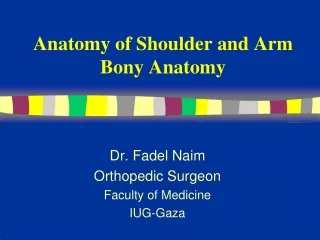
Anatomy of Shoulder Complex. Dr. Fadel Naim Orthopedic Surgeon Faculty of Medicine IUG-Gaza. Trapezius. Origin Medial third superior nuchal line on the occipital bone External occipital protuberance Ligament nuchae Spine of 7 th cervical vertebra

E N D
Anatomy of Shoulder Complex Dr. FadelNaim Orthopedic Surgeon Faculty of Medicine IUG-Gaza
Trapezius • Origin • Medial third superior nuchal line on the occipital bone • External occipital protuberance • Ligament nuchae • Spine of 7th cervical vertebra • Spinous processes and supraspinous ligaments to T12 • INSERTION • Upper fibers to lateral third of posterior border of clavicle • Middle fibers to medial acromion and superior lip of spine of scapula • Lower fibers to medial end of spine of scapula • Action • Laterally rotates, elevates and retracts scapula. • If scapula is fixed, extends and laterally flexes neck • Nerve • Motor fibers form spinal accessory nerve (Cranial nerve XI) • Sensory fibers spinal nerves C3 and C4
Levator Scapulae • Origin • Posterior tubercles of transverse processes of C1-4 • INSERTION • Upper part of medial border of scapula opposite the supraspinous fossa • Action • Raises medial border of scapula • In conjunction with middle fibers of trapezius and rhomboids, pulls the scapula medially and upward • Nerve • Anterior primary rami of C3 and C4 and dorsal scapular nerve (C5)
Rhomboid Minor • Origin • Lower ligamentum nuchea • Spines of C7 and T1 • INSERTION • Small area of posteromedial border of scapula at level of spine, below levator scapulae • Action • Elevates the medial border of the scapula and pulls it medially • Nerve • Dorsal scapular nerve (C5)
Rhomboid Major • Origin • Spines of T2-T5 and supraspinous ligaments • INSERTION • Lower half of posteromedial border of scapula, opposite the infraspinous fossa • Action • Elevates the medial border of the scapula and pulls it medially • Nerve • Dorsal scapular nerve (C5) (from root )
Deltoid • ORIGIN • Anterior fibers: Lateral third of clavicle • Middle fibers: Acromion • Posterior fibers: spine of scapula to deltoid tubercle • INSERTION • Deltoid tuberosity • Middle of lateral surface of humerus • ACTION • Abducts arm • anterior fibers flex and medial rotate • posterior fibers extend and lateral rotate • NERVE • Axillary nerve (C5, 6) (from posterior cord) Forms the rounded contour of the shoulder
Supraspinatus • Origin • Medial three quarters of supraspinous fossa of scapula • Upper surface of spine • INSERTION • Superior facet on greater tuberosity of humerus • Capsule of shoulder joint • Action • Abducts arm • Stabilizes shoulder joint • Nerve • Suprascapular nerve
Infraspinatus • Origin • Medial three quarters of infraspinous fossa of scapula • INSERTION • Middle facet of greater tuberosity of humerus • Capsule of shoulder joint • Action • Laterally rotates arm • Stabilizes shoulder joint • Nerve • Suprascapular nerve
Teres Minor • Origin • Middle third lateral border of scapula above teres major • INSERTION • Inferior facet of greater tuberosity of humerus (below infraspinatus) • Capsule of shoulder joint • Action • Laterally rotates arm • Stabilizes shoulder joint • Nerve • Axillary nerve
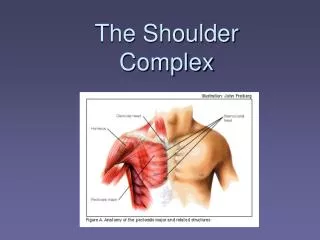
Pectoralis Minor • Origin 3, 4, 5 ribs • INSERTION • Medial and upper surface of coracoid process of scapula • Action • Elevates ribs if scapula fixed • Pulls shoulder downward and forward • Nerve • Medial pectoral nerve (from medial cord of brachial plexus)
Quadrangular space • It is bounded: • Superiorly by: • Subscapularis • Teres minor • Inferiorly by: • Teres major • Medially by: • Long head of triceps • Laterally by: • Humerus The axillary nerve and the posterior circumflex humeral vessels pass backward through this space
Triangular space • an area of communication between the axilla and the posterior scapular region • formed by: • the medial margin of the long head of triceps brachii • the superior margin of teres major • the inferior margin of teres minor The circumflex scapular artery and vein pass through this gap
Triangular interval • formed by: • the lateral margin of the long head of triceps brachii; • the shaft of the humerus; • the inferior margin of teres major The radial nerve, the profundabrachii artery and associated veins pass through it
Quadrangular space syndrome • Hypertrophy of the quadrangular space muscles or fibrosis of the muscle edges may impinge on the axillary nerve. • Uncommonly, this produces weakness of the deltoid muscle. • Typically it produces atrophy of the teres minor muscle • which may affect the control that the rotator cuff muscles exert upon shoulder movement
Rotator Cuff Muscles • Four of the scapulohumeral muscles • Supraspinatus • Infraspinatus • Teres minor • Subscapularis • Referred to as SITS muscles • Called rotator cuff muscles because they form a musculotendinous cuff around the glenohumeral joint {Dynamic stabilizers (“cuff”)} • All except the supraspinatusare rotators of the humerus
The tendons of the muscle blend with the fibrous capsule of the glenohumeral joint to form a musculotendinous rotator cuff, which reinforces the capsule on three sides: • Anteriorly • Superiorly • Posteriorly • The cuff is weakest antero-inferiorly, making shoulder dislocation most common in this direction.
Injury to rotator cuff can occur in any of the following manners: • Acutely, following trauma; • As a result of chronic impingement and overuse (excessive abduction); • Compromised blood supply. • The most frequently injured tendon is the supraspinatus, probably because it is relatively avascular • Additionally, the supraspinatus tendon is subject to significant trauma as it is compressed between the acromion and the humeral head during abduction
Types Of Acromion • Three distinct types of acromion can be seen on the angled outlet Y view: • The type I acromion, which is flat, is the "normal" acromion. • The type II acromion is more curved and downward dipping, • the type III acromion is hooked and downward dipping, obstructing the outlet for the supraspinatus tendon. • Cadaveric studies have shown an increased incidence of rotator cuff tears in persons with type II and type III acromions
Rotator Cuff Injuries • Injury or disease may damage the musculotendinous rotator cuff, producing instability of the glenohumeral joint • Trauma may tear or rupture one or more of the tendons of the muscles forming the rotator cuff • Acute tears may occur when the arm is violently pushed into abduction • The patient reports a sharp pain in the anterosuperior part of the shoulder • Rotator cuff injuries are also common in persons with throwing activities • Rotator cuff tears also follow dislocation of the shoulder
Degenerative tendonitis of the rotator cuff is common, especially in old people. • To test for this disease, the person is asked to lower the fully abducted limb slowly and smoothly. • From an approximately 90 ° abduction, the limb will suddenly drop to the side in an uncontrolled manner if the rotator cuff (especially the supraspinatus part) is diseased and torn • The injury often results from an indirect force to the abducted arm, such as a fall in a person older than 45 years • Acute tears are uncommon in young persons.
This injury causes tenderness around the greater tubercle of the humerus and pain during 45 ° of passive abduction. • X-rays may be normal, but can reveal: • Narrowing of the distance between the acromion and humeral head (impingement) • Erosion of the inferior acromion • Sclerosis of the greater tuberosity
130° Subacromial Bursitis 50° • The tendon of the supraspinatus is separated from the coracoacromial ligament, acromion, and deltoid by the subacromial bursa. • Painful arc syndrome • When this bursa is inflamed abduction of the arm is extremely painful during the arc of 50 to 130 °. • The pain may radiate as far distally as the hand. • Acute pain is also felt lateral to the acromion. • A painful arc signifies an impingement of a painful structure during the movement of the humerus in relation to the roof of the shoulder
The commonnest pathological situations causing painful arc are: • 1. Tendinitis of Supraspinatus • 2. Tendinitis of Infraspinatus • 3. Tendinitis of Subscapularis • 4. Tendinitis of the long head of the Biceps • 5. Subacromial Bursitis • 6. AC Sprain • Less frequent pathologies associated with a painful arc are: • metastases in the head of the humerus • metastases in the acromion • Instability of the shoulder
Scapular Motions Rotational Elevation Of Glenoid Cavity Rotational Depression Of Glenoid Cavity
The supraspinatus initiates abduction and the deltoid takes over once the humerus is abducted past 15 degrees • During full abduction of the arm we have to laterally rotate our humerus to move the greater tuberosity out of the way. • Even with this maneuver, space is too limited to allow for the range of motion that is seen in a normal individual. • The remainder of the range of motion is made possible by scapular rotation. • Once we have abducted the arm past 20 degrees or 30 degrees, for every 3 degrees of abduction at the glenohumeral joint, 1 degree occurs at the scapulothoracic surface and only 2 degrees occurs at the glenohumeral joint.