Download
1 / 45
651 likes | 1.23k Views
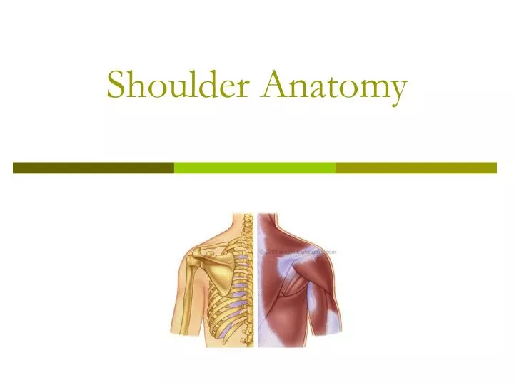
Shoulder Anatomy. Shoulder. It is a ball and socket joint that moves in all three planes and has. Most mobile and least stable joint. Shoulder joint motions. Flexion- is raising the arm in the lateral plane from 0-180 degrees. Extension- return to anatomical position.

E N D
Shoulder • It is a ball and socket joint that moves in all three planes and has. • Most mobile and least stable joint.
Shoulder joint motions • Flexion- is raising the arm in the lateral plane from 0-180 degrees. • Extension- return to anatomical position. • Hyperextension- 0-45 degrees back through the lateral plane.
Shoulder joint motions • Abduction- arm moving in the frontal plane away from the body, with a 0-180 degrees of motion. • Adduction- arm moving back to midline, with 0-180 degrees of motion.
Internal Rotation- occur in the transverse plane. This can go to 90 degrees into body External Rotation- occurs in the transverse plane, 90 degrees out from neutral. Shoulder joint motions
Horizontal abduction/adduction- occurs in the transverse plane. Neutral is 90 degrees of shoulder abduction, so horiz abduction is 30 degrees and adduction is 120 degree. Shoulder joint motions
Scaption- flexion in the scapular plane, vs the lateral or frontal plane. 180 degree of motion can occur. Shoulder joint motions
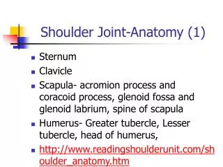
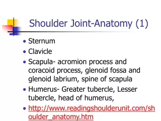
Shoulder Landmarks Scapula Glenoid labrum-fibrocartilage ring attached to the rim of the glenoid fossa, which deepens the cavity.
Humerus- Head- is the semi round proximal end, articulates with the scapula. Shaft- body of the humerus is the area between the neck and the epicondyles. Shoulder Landmarks
Surgical Neck- where the head meets the body. Anatomical neck- where the head meets the tubercles. Shoulder Landmarks
Shoulder Landmarks Greater Tubercle/Tuberosity- large projection lateral to the head. Supraspinatus, infraspinatus and teres minor attach here.
Shoulder Landmarks Lesser Tubercle/Tuberosity- smaller projection on the anterior surface, subscapularis attaches here.
Shoulder Landmarks Deltoid tuberosity- lateral side, near the midpoint, deltoid attaches here.
Shoulder Landmarks Bicipital Groove- groove between the tubercles containing the long head of the biceps tendon.
Impingement Syndrome • A condition that occurs when the space between the humeral head and the acromion above becomes narrowed. • The three things that can get pinched are the: joint capsule, tendons of rotator cuff, and bursa.
Impingement Syndrome • Impingement can create either bursitis, or tendonitis depending on what structure is being squeezed. • Overhead athletes are more likely to have problems with this injury. • 1/3 of shoulder problems are due to impingement.
Signs and Sx Pain and tender GH joint Pain and weak active abd in mid range Limited internal rotation + Hawkins Test Tender subacromial area possibly into the deltoid Treatment Correct technique Strengthen inferior muscles Strengthen weak rotator cuff muscles Impingement Syndrome
Impingement Syndrome • Special Tests • Hawkins Test • Neer’s Impingement • Cross over Test
Impingement Syndrome • Stretches- • 3 way door stretch • Posterior shoulder • Internal Rotation with • Exercises • Internal Rotation • External Rotation • Adduction
Rotator Cuff Tears • In the young person it is more of a traumatic injury, fall on outstretched arm, arm yanked back. • Young person can have chronic injury that ultimately tears a tendon. • In the older person it is a result of lose of elasticity in the muscle and tendon and can tear with everyday activities or a bone spur.
Rotator Cuff Tears • Signs and Sx • With a parcial tear the athlete will feel pain but still be able to move with normal ROM. • With a complete tear the athlete will not have normal ROM. • Overhead motions are hardest. • A shrug motion will result. • Pain sleeping on injured side.
Rotator Cuff Tears • Special Tests • Active Abduction-look for hiking shoulder • Drop Arm sign- athlete abduct above head then lowers slow, look for loss of muscle control. • Supraspinatus muscle test- looking for weakness • Empty Can Test- supraspinatus/subscap motion • MRI is final diagnostic tool
Biceps Tendonitis • Discomfort in the front of the shoulder. • Can be caused by impingement. • Special Tests- • Speed’s Test • Yergeson’s Test
Traumatic Shoulder Injuries • Shoulder Dislocation • Glenoid Labrum Injuries • Multidirectional Instabilites • Acromioclavicular Separation • Brachial Plexus Injury • Fractures
Anterior Shoulder Dislocation • A humerus can dislocate • Anteroinferiorly-front and down (most common) • Inferiorly – down • Posteriorly -back
Anterior Shoulder Dislocation • Anterior dislocation happens when the arm is abducted to the side and a forceful external rotation happens. • A doctor visit is necessary, immediately if the humerus does not relocate on it’s own. • Even if it goes back a Hill-Sach’s Lesion can occur.
Anterior Shoulder Dislocation • Rehabilitation is very important to this injury. • Reinjury will likely happen if a first time injury happens before the age of 20. • Surgery may be necessary if repeated dislocation occurs.
Special Test-Dislocation • Apprehension test
Glenoid Labrum Injury • Glenoid Labrum-a ring of cartilage attached to the margin of the glenoid cavity of the scapula. • The labrum acts to keep the humeral head positioned on the glenoid by blocking unwanted movement.
Glenoid Labrum Injury • A labral tear can occur with a shoulder dislocation, more likely to occur with numerus dislocations. • A degenerative tear can occur when a shoulder becomes loose, letting the humeral head slip over the labrum numerus times and eventually the labrum will fail/tear.
Signs and Sx Pain with catching and popping Possible weakness Possible limited ROM Special Tests Clunk Test Cross Over Test Treatment Rotator Cuff strengthening Surgery Glenoid Labrum Injury
Multidirectional Instabilities • Typically an anatomical problem. • Multiple dislocations will make it worse. • Exercise may help with the problem, surgery sometimes, but not always • Weight bearing exercise are helpful. Like what?
Acromicavicular Separation • Also known as an AC sprain. • Occurs due to fall on outstretched arm or tip of shoulder. May be due to blow to tip of shoulder
Signs and Sx deformity Pain in vicinity of AC Special Test Shear Test Sulcus Sign Treatment Three grades –the grade determines treatment Grade one is exercise and ice Grade two immobilize 3 weeks and then exercise Grade three immobilize 5 weeks and then exerccise AC separation
Muscles of the Shoulder Joint • Deltoid is superficial muscle. All three parts of it attach to the deltoid tuberosity. • Axillary Nerve
Rotator Cuff Muscles • Supraspinatus-anterior superior shoulder. It is superior to the spine of the scapula. • abduction
Muscles of the Shoulder Joint • Pectoralis Major- • Clavicular portion-most effective during flexion from 0-90 • Sternal portion- most effective in extension 180-120 degrees of shoulder extension • Both of them adduct, internally rotate and horizontally adduct the shoulder.
Latissimus Dorsi- means widest, back, so the widest back muscle. It is mostly superficial and is involved with shoulder extension , adduction and internal rotation Muscles of the Shoulder Joint
Teres Major- it is the little helper of the lats. It runs from the axillary boarder of the scapula to the lesser tubercle of the humerus. Muscles of the Shoulder Joint
Rotator Cuff Muscles • Infraspinatus- • posterior inferior shoulder • Inferior to the spine of the scapula • External rotation
Rotator Cuff Muscles • Teres Minor- posterior shoulder • Adduction
Rotator Cuff Muscles • Subscapularis-anterior shoulder • Internal rotation
Muscles of the shoulder joint • Coracobrachialis- attaches to the coracoid process and the arm or Brachium. Stabalizes the humerus in the fossa.
The four rotator cuff muscles cover the humeral head and hold the head against the glenoid fossa. Muscles of the Shoulder Joint
Rotator Cuff Muscles • Know these muscles if you remember nothing else. • Infraspinatus • Supraspinatus • Subscapularis • Teres Minor